

Redefining POCUS with Accessible, Standardized Imaging
In the rapidly evolving landscape of medical imaging technology, portable ultrasound (POCUS ultrasound) has emerged as a game-changer, enabling healthcare providers to deliver timely, accurate diagnostics at the point of care. As a global leader in ultrasound innovation, SonoStar (Sonostar Group) is set to revolutionize the market once again with the upcoming launch of its new series of 3D wireless handheld ultrasound devices. This groundbreaking lineup builds on the success of traditional 2D handheld ultrasound, integrating advanced 3D imaging capabilities while maintaining ultra-low costs—only slightly higher than conventional 2D portable ultrasound systems. Designed to support a wide range of transducers, including convex array, linear array (equipped with electronic array scanning, mechanical swing scanning, and rotational scanning), and covering a frequency range of 2MHz to 20MHz, the new 3D wireless handheld ultrasound from SonoStar is poised to transform clinical practice across obstetrics, aesthetic medicine, thyroid and breast imaging, carotid artery screening, musculoskeletal (MSK) diagnostics, and more.
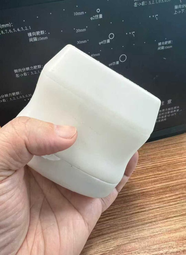
SONOSTARGROUP 3D Probe
SONOSTARGROUP 3D Probe
SONOSTARGROUP 3D Probe
The rise of POCUS ultrasound (Point-of-Care Ultrasound) has underscored the need for compact, portable, and user-friendly imaging solutions that do not compromise on diagnostic accuracy. Traditional ultrasound systems, while powerful, are often large, expensive, and confined to dedicated imaging suites, limiting their accessibility in primary care clinics, remote medical facilities, emergency settings, and even mobile healthcare units. Handheld ultrasound devices have addressed many of these challenges, but until now, 3D imaging—a technology that provides volumetric, comprehensive views of anatomical structures—has been largely reserved for high-end, stationary ultrasound systems. SonoStar’s new 3D wireless handheld ultrasound bridges this gap, bringing clinic-grade 3D imaging to the palm of the hand, supported by a wireless probe design that eliminates the constraints of cumbersome cables and enhances mobility.
Core Product Highlights: 3D Imaging, Versatile Transducers, and Unbeatable Cost-Effectiveness
At the heart of SonoStar’s upcoming 3D wireless handheld ultrasound series is its commitment to accessibility, versatility, and performance. Unlike many 3D ultrasound systems that come with prohibitive price tags, SonoStar has engineered this new lineup to be cost-competitive, with a price point only marginally higher than 2D handheld ultrasound devices. This cost advantage is a critical breakthrough, as it enables smaller clinics, community health centers, and healthcare providers in resource-constrained regions to adopt 3D imaging technology—previously a luxury for large hospitals and specialized imaging centers. For healthcare organizations looking to upgrade their ultrasound capabilities without overstretching their budgets, this 3D wireless handheld ultrasound represents an ideal solution, balancing advanced features with economic feasibility.
A key differentiator of the SonoStar 3D wireless handheld ultrasound is its support for multiple transducer types, including convex array and linear array probes. Convex array ultrasound probes are widely used for abdominal, pelvic, and obstetric imaging, thanks to their curved design that allows for a wider field of view and deeper penetration—critical for visualizing large organs and fetal structures. Linear array ultrasound probes, on the other hand, are ideal for superficial imaging, such as thyroid, breast, carotid artery, and musculoskeletal scans, as they deliver high-resolution images of structures close to the skin’s surface. What sets SonoStar’s offering apart is its integration of dual scanning technologies in linear array probes: electronic array scanning, mechanical swing scanning, and rotational scanning. Electronic array scanning uses a matrix of piezoelectric elements to generate 2D and 3D images with high speed and precision, while mechanical swing scanning and rotational scanning enhance volumetric data collection, ensuring comprehensive coverage of the target area. This combination of scanning technologies ensures that the wireless probe can adapt to diverse clinical needs, making it a versatile tool for multi-specialty practices.
The frequency range of 2MHz to 20MHz further amplifies the versatility of the SonoStar 3D wireless handheld ultrasound. Lower frequencies (2MHz to 5MHz) are ideal for deep-tissue imaging, such as abdominal organs, pelvic structures, and fetal imaging in later trimesters, as they penetrate deeper into the body with minimal signal attenuation. Higher frequencies (10MHz to 20MHz) deliver ultra-high resolution for superficial structures, making them perfect for thyroid, breast, carotid artery, and musculoskeletal imaging—where detail is critical for detecting small lesions, nodules, or abnormalities. The ability to switch between low and high frequencies seamlessly, combined with the 3D imaging capability, ensures that healthcare providers can obtain comprehensive, high-quality images across a wide range of clinical scenarios, all with a single handheld device and wireless probe.
Wireless connectivity is another cornerstone of SonoStar’s new 3D handheld ultrasound. The wireless probe design eliminates the need for bulky cables, allowing healthcare providers to move freely around the patient, adjust their position for optimal imaging, and even perform scans in tight spaces—such as emergency vehicles, patient bedsides, or remote medical camps. This mobility is particularly valuable for POCUS ultrasound, where quick, on-the-spot diagnostics can significantly improve patient outcomes. The wireless connection is engineered to be stable and secure, ensuring that image transmission is fast and uninterrupted, with no loss of image quality. Healthcare providers can also easily share 3D ultrasound images with colleagues for consultation, store data in electronic health records (EHRs), or even stream live images to a central monitoring station—enhancing collaboration and workflow efficiency.
3D Ultrasound Imaging: Transforming Clinical Applications Across Specialties
3D ultrasound imaging has long been recognized for its ability to provide volumetric, three-dimensional views of anatomical structures, offering a more comprehensive understanding of patient anatomy than traditional 2D ultrasound. While 2D ultrasound delivers flat, cross-sectional images that require the operator to mentally reconstruct the 3D structure, 3D ultrasound captures volumetric data that can be rotated, sliced, and analyzed from multiple angles—reducing the cognitive load on the operator and improving diagnostic accuracy. SonoStar’s 3D wireless handheld ultrasound makes this advanced technology accessible across a wide range of clinical specialties, from obstetrics to aesthetic medicine, and from primary care to specialized diagnostics.
Obstetric Ultrasound: Enhancing Fetal Monitoring and Prenatal Care
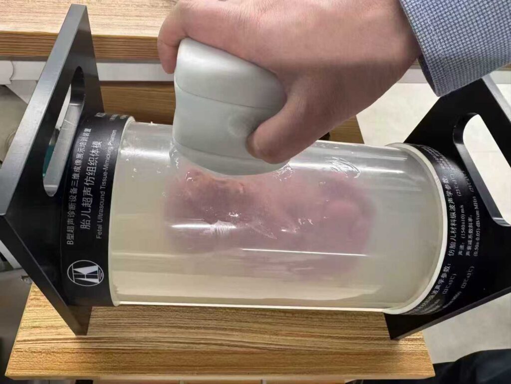
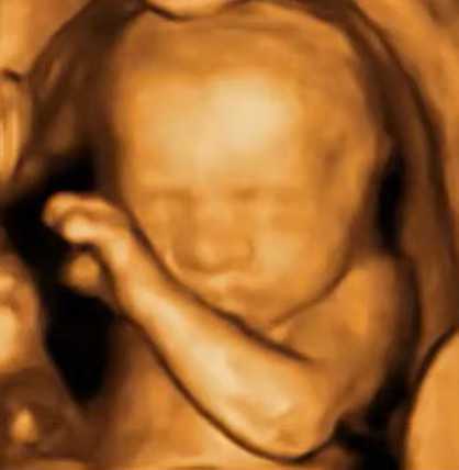
Obstetric ultrasound is one of the most common applications of ultrasound imaging, and 3D technology has revolutionized prenatal care by providing detailed views of the fetus, placenta, and uterine structures. SonoStar’s 3D wireless handheld ultrasound is set to become an indispensable tool for obstetricians, midwives, and prenatal care providers, enabling them to perform comprehensive fetal assessments with a portable, easy-to-use device. The 3D imaging capability allows for clear visualization of fetal facial features, limbs, organs, and fetal position—helping to detect congenital anomalies, monitor fetal growth, and assess placental location and function.
Unlike traditional 2D obstetric ultrasound, which requires the operator to capture multiple cross-sectional images and piece them together, 3D ultrasound captures a complete volumetric dataset in a single scan, reducing the time required for each examination. The wireless probe design allows the provider to move freely around the patient, making it easier to obtain optimal images of the fetus, even in cases where the patient’s position is limited. Additionally, the low cost of SonoStar’s 3D wireless handheld ultrasound makes it accessible to prenatal care clinics in rural and underserved areas, where access to high-end ultrasound systems is often limited. This accessibility can significantly improve prenatal care outcomes, as early detection of fetal anomalies and complications allows for timely intervention and management.
For expectant parents, 3D ultrasound images also provide a more personal connection to the fetus, as they can see detailed, lifelike views of their baby. This emotional benefit, combined with the clinical advantages of 3D imaging, makes SonoStar’s 3D wireless handheld ultrasound a valuable tool for enhancing patient satisfaction in obstetric care.
Aesthetic Medicine Ultrasound: Precision in Filler and Tissue Assessment
The field of aesthetic medicine has seen rapid growth in recent years, with an increasing number of patients seeking non-invasive procedures such as dermal fillers, botulinum toxin injections, and tissue rejuvenation. Ultrasound imaging has become an essential tool in aesthetic medicine, as it allows providers to visualize facial anatomy, including blood vessels, nerves, and soft tissues, ensuring precise placement of fillers and avoiding complications. SonoStar’s 3D wireless handheld ultrasound takes this a step further, providing volumetric views of facial structures that enable even greater precision and safety.
In aesthetic medicine, the ability to visualize the depth and distribution of dermal fillers is critical for achieving natural-looking results and preventing adverse events such as vascular occlusion. 2D ultrasound can provide cross-sectional views of filler placement, but 3D ultrasound allows providers to assess the entire volume of the filler, ensuring that it is evenly distributed and placed in the correct anatomical plane. The high-frequency linear array wireless probe (10MHz to 20MHz) delivers ultra-high resolution images of superficial facial structures, making it easy to distinguish between filler material, soft tissue, and blood vessels.
The wireless and portable design of SonoStar’s 3D handheld ultrasound is particularly well-suited for aesthetic clinics, where space is often at a premium and providers need to move between treatment rooms. The device can be easily carried from room to room, and the wireless probe allows for flexible positioning during scans—critical for imaging different areas of the face and neck. Additionally, the standardized scanning capability of the 3D technology ensures that even less experienced providers can obtain consistent, accurate images, reducing the risk of errors and improving patient safety.
Thyroid and Breast Ultrasound: Early Detection of Abnormalities
Thyroid and breast ultrasound are key diagnostic tools for the early detection of nodules, cysts, and malignancies. These superficial structures require high-resolution imaging, and 3D ultrasound has been shown to improve the characterization of small lesions, making it easier to distinguish between benign and malignant masses. SonoStar’s 3D wireless handheld ultrasound, with its high-frequency linear array wireless probe and 3D imaging capability, is ideally suited for thyroid and breast imaging in both primary care and specialist settings.
For thyroid ultrasound, 3D imaging allows providers to visualize the entire thyroid gland, including the right and left lobes and the isthmus, in a single volumetric scan. This comprehensive view makes it easier to detect small nodules (as small as 2-3mm) and assess their size, shape, margin, and internal echogenicity—key features for determining the risk of malignancy. The wireless probe design allows for easy access to the neck area, and the portable nature of the device means that thyroid scans can be performed in primary care clinics, eliminating the need for patients to wait for referrals to specialized imaging centers.
In breast ultrasound, 3D imaging is particularly valuable for women with dense breast tissue, where mammography may be less effective at detecting small lesions. The 3D wireless handheld ultrasound can be used to perform targeted scans of suspicious areas identified on mammography or clinical exam, providing detailed volumetric images that help to characterize the lesion. The ability to rotate and slice the 3D dataset allows providers to view the lesion from multiple angles, improving the accuracy of diagnosis and reducing the need for unnecessary biopsies.
Carotid Artery Ultrasound: Screening for Vascular Disease
Carotid artery disease, characterized by the buildup of plaque in the carotid arteries, is a major risk factor for stroke. Early screening and detection of carotid artery stenosis (narrowing of the arteries) are critical for preventing stroke and improving patient outcomes. Carotid artery ultrasound is a non-invasive, cost-effective screening tool that uses ultrasound waves to visualize the carotid arteries and measure blood flow velocity. SonoStar’s 3D wireless handheld ultrasound enhances carotid artery screening by providing 3D volumetric images of the carotid arteries, allowing for more accurate assessment of plaque burden and stenosis.
Traditional 2D carotid artery ultrasound requires the operator to capture multiple cross-sectional images of the common carotid artery, internal carotid artery, and external carotid artery, as well as measure blood flow velocity using Doppler ultrasound. 3D ultrasound simplifies this process by capturing a complete volumetric dataset of the carotid arteries, which can be analyzed to measure the degree of stenosis, assess plaque morphology (e.g., calcified vs. soft plaque), and evaluate blood flow dynamics. The wireless probe design allows for easy positioning around the neck, and the portable nature of the device makes it ideal for mobile screening programs, such as community health fairs or senior care facilities.
The low cost of SonoStar’s 3D wireless handheld ultrasound also makes it accessible to primary care clinics and rural healthcare facilities, where carotid artery screening is often underutilized due to the high cost of traditional ultrasound systems. By making carotid artery screening more accessible, SonoStar’s device has the potential to reduce the incidence of stroke and improve public health outcomes.
Musculoskeletal (MSK) Ultrasound: Diagnosing and Managing Orthopedic Conditions
Musculoskeletal (MSK) ultrasound is used to diagnose and manage a wide range of orthopedic conditions, including tendonitis, bursitis, ligament tears, muscle strains, and joint effusions. MSK ultrasound requires high-resolution imaging of superficial structures (tendons, ligaments, muscles, and joints) and often requires dynamic scanning (imaging while the patient moves the affected limb). SonoStar’s 3D wireless handheld ultrasound is ideally suited for MSK imaging, thanks to its high-frequency linear array wireless probe, 3D imaging capability, and wireless mobility.
The 3D imaging capability of SonoStar’s device allows for comprehensive visualization of MSK structures, including the entire length of a tendon or ligament, in a single volumetric scan. This makes it easier to detect tears, inflammation, or degenerative changes that may not be visible on 2D ultrasound. The wireless probe design allows the provider to move freely around the patient, making it easy to perform dynamic scans—critical for assessing the function of tendons and ligaments during movement. For example, when evaluating a patient with a suspected Achilles tendon tear, the provider can scan the tendon while the patient plantarflexes and dorsiflexes the foot, allowing for real-time assessment of tendon integrity.
The portability of the 3D wireless handheld ultrasound also makes it valuable for orthopedic surgeons, sports medicine physicians, and physical therapists who need to perform on-the-spot diagnostics in clinics, training rooms, or even at the bedside. The device can be used to guide minimally invasive procedures, such as steroid injections or ultrasound-guided aspirations, ensuring precise placement of the needle and reducing the risk of complications.
Standardized Scanning: Eliminating Operator Dependence and Improving Diagnostic Consistency
One of the most significant challenges in ultrasound imaging—especially in POCUS ultrasound—is the dependence on operator skill. Traditional 2D ultrasound requires extensive training and experience to obtain high-quality images, as the operator must know how to position the probe, adjust the settings, and capture the correct cross-sectional views. Variability in operator technique can lead to inconsistent image quality, missed abnormalities, and misdiagnoses—particularly in settings where less experienced providers perform ultrasound scans.
SonoStar’s 3D wireless handheld ultrasound addresses this challenge by introducing standardized scanning capabilities that reduce operator dependence and improve diagnostic consistency. Unlike 2D ultrasound, which relies on the operator to capture individual cross-sectional images, 3D ultrasound captures a complete volumetric dataset of the target area with a single scan. This means that the operator does not need to have advanced probe manipulation skills to obtain high-quality images—they simply position the wireless probe over the target area, and the device automatically captures the entire volume of data.
The standardized scanning feature of SonoStar’s 3D wireless handheld ultrasound has several key benefits. First, it lowers the barrier to entry for ultrasound imaging, allowing less experienced providers (such as primary care physicians, nurses, and emergency medical technicians) to perform accurate ultrasound scans without extensive training. This is particularly valuable in POCUS ultrasound, where timely diagnostics are critical and access to experienced ultrasound technicians may be limited. Second, it reduces variability in image quality and diagnostic results, ensuring that patients receive consistent care regardless of which provider performs the scan. Third, it improves efficiency by reducing the time required to perform each scan—there is no need to repeat scans due to poor image quality, and the volumetric dataset can be analyzed later if additional views are needed.
In addition to standardized scanning, SonoStar’s 3D wireless handheld ultrasound is equipped with advanced image processing algorithms that automatically optimize image quality, adjust the frequency, and enhance contrast—further reducing the need for operator intervention. The device also features user-friendly software that allows providers to easily navigate, rotate, and slice the 3D dataset, making it easy to analyze the images and make a diagnosis.
The Market Impact of SonoStar’s 3D Wireless Handheld Ultrasound
The global POCUS ultrasound market is growing rapidly, driven by increasing demand for portable, accessible imaging solutions, rising prevalence of chronic diseases, and growing adoption of ultrasound in primary care and emergency settings. According to industry reports, the global POCUS ultrasound market is expected to reach $XX billion by 2030, with a compound annual growth rate (CAGR) of XX%. SonoStar’s 3D wireless handheld ultrasound is well-positioned to capture a significant share of this growing market, thanks to its unique combination of 3D imaging capabilities, versatile transducer support, low cost, and standardized scanning.
One of the key market differentiators for SonoStar’s device is its cost-effectiveness. By offering 3D imaging at a price point only slightly higher than 2D handheld ultrasound, SonoStar is making advanced imaging technology accessible to a broader range of healthcare providers—including those in resource-constrained regions. This accessibility can help to address healthcare disparities by ensuring that patients in rural and underserved areas have access to the same high-quality diagnostic tools as those in urban centers.
Another key market advantage is the device’s versatility. By supporting multiple transducer types (convex array, linear array) and scanning technologies (electronic array, mechanical swing, rotational), and covering a wide frequency range (2MHz to 20MHz), the 3D wireless handheld ultrasound can be used across multiple clinical specialties. This versatility makes it an attractive investment for healthcare organizations that want to upgrade their ultrasound capabilities without purchasing multiple devices for different specialties.
The wireless and portable design of the device also aligns with the growing trend toward mobile and remote healthcare. As telemedicine and mobile health (mHealth) continue to expand, there is increasing demand for portable diagnostic tools that can be used outside of traditional healthcare settings. SonoStar’s 3D wireless handheld ultrasound is ideal for telemedicine applications, as it allows providers to perform scans remotely and share 3D images with specialists for consultation—improving access to care for patients in remote areas.
Conclusion: SonoStar’s 3D Wireless Handheld Ultrasound—The Future of POCUS
SonoStar’s upcoming series of 3D wireless handheld ultrasound devices represents a significant advancement in POCUS ultrasound technology. By combining 3D imaging capabilities, versatile transducer support, a wide frequency range, and ultra-low cost, this groundbreaking device is set to redefine the standards of portable ultrasound imaging. Whether used in obstetrics, aesthetic medicine, thyroid and breast imaging, carotid artery screening, musculoskeletal diagnostics, or other clinical specialties, the 3D wireless handheld ultrasound from SonoStar offers healthcare providers a powerful, accessible, and user-friendly tool for delivering high-quality, timely diagnostics at the point of care.
The standardized scanning feature of the device eliminates operator dependence, ensuring consistent image quality and diagnostic results—even for less experienced providers. The wireless probe design enhances mobility and flexibility, making it ideal for use in a wide range of clinical settings, from primary care clinics and emergency rooms to remote medical facilities and mobile healthcare units. And with a price point only slightly higher than traditional 2D handheld ultrasound, SonoStar’s 3D wireless handheld ultrasound is poised to make advanced 3D imaging accessible to healthcare providers around the world, improving patient outcomes and addressing healthcare disparities.
As the global demand for POCUS ultrasound continues to grow, SonoStar remains committed to innovation, accessibility, and quality. The launch of the 3D wireless handheld ultrasound series is a testament to this commitment, and it is sure to solidify SonoStar’s position as a leader in the global ultrasound market. Healthcare providers, clinics, and hospitals looking to upgrade their ultrasound capabilities and deliver better patient care should keep a close eye on this revolutionary device—set to launch soon and transform the future of point-of-care ultrasound.
For more information about SonoStar’s 3D wireless handheld ultrasound, its features, applications, and availability, visit the official SonoStar Group website at sonostargroup.com. Discover how this advanced ultrasound technology can enhance your clinical practice, improve diagnostic accuracy, and deliver better patient outcomes—all with the convenience of a portable, wireless device.
Leave a Reply