
Description
1. Core Scanning & Probe Parameters
Core hardware and scanning specifications that ensure precise rectal and perianal tissue imaging:
-
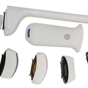
Scanning Mode: Electronic array scanning with biplane rectal probe design, enabling vertical and transverse cross-sectional imaging for comprehensive analysis of rectal wall layers, perianal structures, and lesion relationships.
-
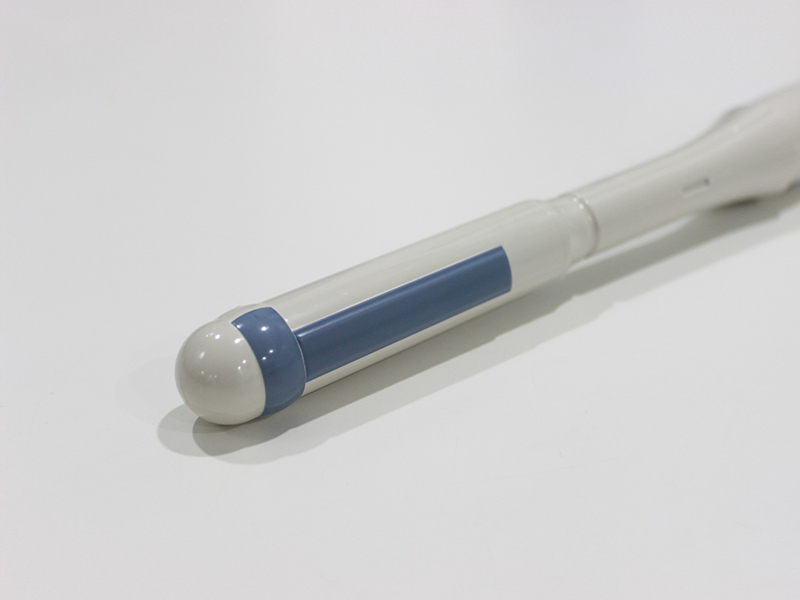
Probe Specifications: Dual-frequency adjustment (6.5MHz/8MHz) – 6.5MHz for balanced penetration and resolution in deep rectal tissues, 8MHz for clear display of superficial perianal structures and subtle lesions.
-
Imaging Range: Adjustable scan depth (40-100mm), perfectly matching the depth needs of rectal and pelvic superficial-to-medium tissue examinations, ensuring complete coverage of target areas.
-
Imaging Foundation: 18 frames/second image frame rate, ensuring smooth real-time imaging for dynamic observation of rectal peristalsis and blood flow changes; 256-level image gray scale for accurate reproduction of tissue density differences.
2. Imaging Modes & Optimization Functions
Comprehensive imaging modes and adjustment tools to enhance rectal imaging clarity and diagnostic precision:
-
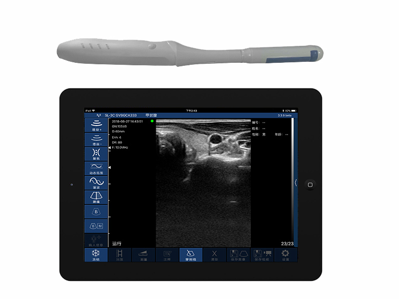
Image Modes: Supports B, B/M, Color Doppler, PW (Pulsed Wave), and PDI (Power Doppler Imaging) modes, covering static tissue morphology observation, dynamic process monitoring, and blood flow signal detection for rectal lesions.
-
Image Adjustment: Equipped with multi-dimensional optimization tools, including Gain (30dB-105dB), 8-level Dynamic Range (40-110), 5-level Noise Reduction (0-4), Focus, Depth, and Harmonic adjustment, effectively reducing image noise and enhancing tissue layering.
-
Cineplay Function: Auto and manual playback with adjustable frames (100/200/500/1000), facilitating frame-by-frame review of key imaging details and accurate lesion characterization.
3. Clinical Auxiliary & Data Management
Practical clinical tools and standardized data handling to improve rectal examination efficiency:
-
Puncture Assist Function: Integrated with in-plane and out-of-plane puncture guide lines, improving the accuracy and safety of perianal and rectal interventional procedures, such as abscess drainage and biopsy.
-
Measurement Functions: Supports Length, Area, Angle, and Obstetric parameters related to pelvic structures, providing accurate quantitative data for evaluating lesion size, tissue thickness, and rectal anatomical relationships.
-
Data Storage: Saves images/videos in JPG, MP4, PNG, and DCM formats; DCM compatibility enables seamless integration with hospital PACS systems, facilitating data archiving and case sharing.
4. Wireless Transmission & Physical Specifications
High-speed wireless transmission and ergonomic design optimized for rectal examination scenarios:
-
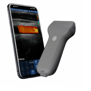
Wireless & Compatibility: 802.11g/20MHz/5G WiFi with 450Mbps transmission speed, enabling stable wireless data transmission; compatible with Android/iOS systems for flexible connection to smartphones/tablets.
-
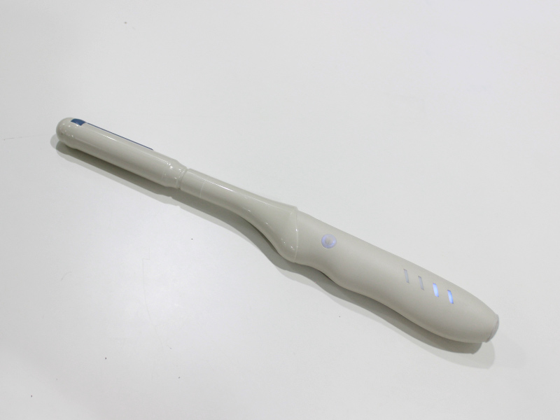
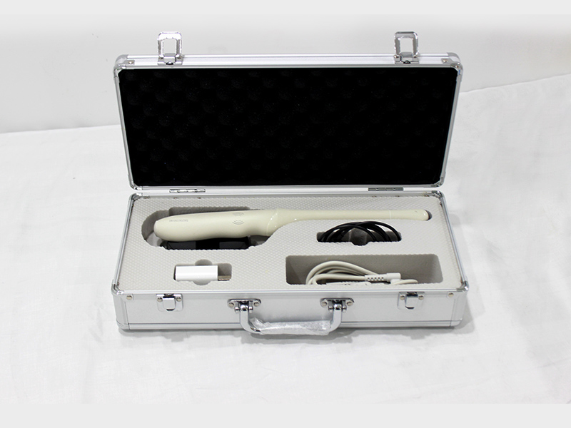
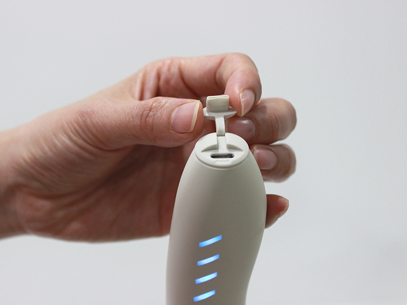
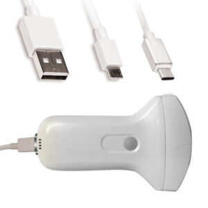
Power Supply: Built-in battery supports 1 hour of continuous operation; USB charging and optional wireless charger ensure sustainable use in clinical settings without power constraints.
-
Physical Dimensions: 336mm×35mm×28mm compact size, 200g net weight, 360mm overall length; ergonomic design for comfortable handheld operation during rectal examinations, easy to carry in medical bags.
The VISION4 CR biplane rectal palm Doppler ultrasound scanner integrates specialized biplane imaging, comprehensive Doppler functions, and flexible wireless transmission. Tailored for rectal and perianal disease diagnosis, it provides clear tissue visualization, precise interventional guidance, and efficient data management. Its compact design and stable performance make it ideal for clinics, specialist departments, and mobile medical services, balancing diagnostic accuracy and operational convenience in anorectal examination scenarios.
Your message has been sent

Reviews
There are no reviews yet.