
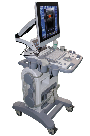
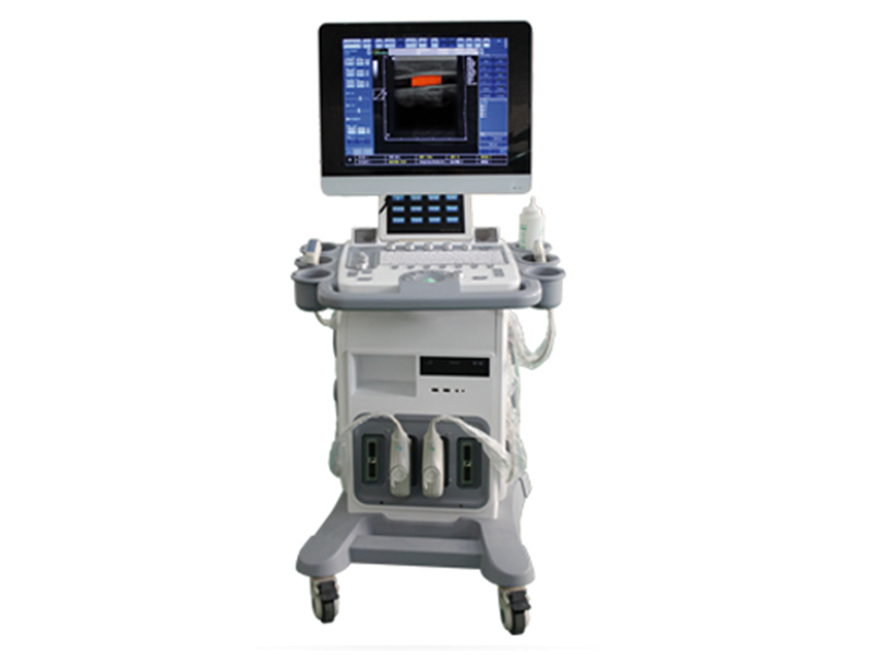
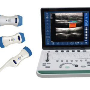
Description
1. Core Product Features
The VISION C200 is engineered with a focus on performance, usability, and adaptability, featuring the following key attributes:
- High-Definition Imaging: Advanced signal processing and tissue harmonic technology deliver crisp, clear images with exceptional detail resolution, enabling accurate detection of subtle anatomical variations and lesions.
- 15-Inch Touchscreen LED Display: A large, responsive touchscreen interface simplifies operation, allowing for intuitive navigation of settings, imaging modes, and measurement tools. The LED backlight ensures bright, consistent visibility across different clinical environments.
- Built-In Battery: Integrated rechargeable battery provides uninterrupted operation during power outages or when mobility is required (e.g., bedside examinations, emergency scenarios), enhancing clinical flexibility.
- Optional 3D/4D Imaging: Available 3D/4D imaging capability offers volumetric visualization for specialized applications such as fetal anatomy assessment, obstetric monitoring, and structural analysis of organs, elevating diagnostic precision.
- Optional Phased Array Probe Support: Compatibility with phased array probes extends the system’s utility to cardiac imaging, enabling detailed evaluation of cardiac structures and function.
- Multilingual & Feature-Rich: Supports multiple languages to cater to diverse healthcare settings, with an extensive suite of imaging modes, measurement tools, and post-processing functions.
- Dual Storage Options: Equipped with a built-in hard disk and USB connectivity for secure, convenient storage of images and videos in multiple formats, facilitating documentation and data sharing.
- Elegant & Ergonomic Design: A sleek, durable trolley design with intuitive component placement reduces operator fatigue, while the system’s robust construction ensures longevity in high-volume clinical environments.
2. Imaging Modes & Clinical Capabilities
The VISION C200 offers a comprehensive range of imaging modes to support diverse diagnostic needs, ensuring versatility across specialties:
- 2D Imaging Modes: Includes B-mode (Brightness mode), B|B (Dual B-mode), 4B (Quad B-mode), B|M (B-mode + M-mode), and M-mode (Motion mode). These foundational modes provide real-time 2D visualization of anatomical structures, motion tracking (M-mode), and simultaneous multi-view imaging (B|B, 4B) for comparative analysis.
- Doppler Imaging Modes:
- Color Doppler Flow Mapping (CFM): Visualizes blood flow direction and velocity in real time, aiding in the detection of vascular abnormalities such as stenosis, occlusion, or abnormal perfusion.
- Power Doppler Imaging (PDI): Enhances sensitivity to low-velocity blood flow, ideal for evaluating microvascular networks in organs, tumors, or peripheral tissues.
- Directional Power Doppler (DPDI): Combines the sensitivity of PDI with directional flow information, improving accuracy in vascular assessment.
- Pulsed Wave Doppler (PWD): Quantifies blood flow velocity with spectral analysis, supporting precise measurement of hemodynamic parameters (e.g., peak velocity, resistance index).
- Duplex (B+PWD) & Triplex (B+CFM/PDI/DPDI+PWD): Integrates 2D imaging with Doppler modalities for simultaneous structural and functional assessment, critical for vascular and cardiac studies.
- Advanced Imaging Enhancements:
- High Pulse Repetition Frequency (HPRF): Optimizes Doppler imaging for high-velocity blood flow (e.g., arterial stenosis), reducing aliasing and improving flow visualization.
- Tissue Harmonic Imaging (THI): Minimizes artifacts and enhances tissue contrast, particularly useful for deep abdominal imaging, obese patients, or regions with poor acoustic window.
3. Scanning Methods & Technical Parameters
3.1 Scanning Flexibility
The system supports multiple scanning methods to adapt to diverse anatomical regions and clinical scenarios:
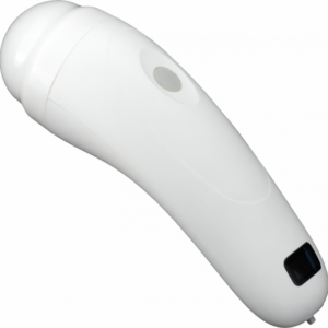
- Scanning Types: Electronic linear (for superficial structures), electronic convex (for abdominal, pelvic, and obstetric imaging), electronic micro-convex (for pediatric, small organ, or interventional use).
- Scanning Depth: Adjustable from 2cm to 24cm, covering superficial (e.g., thyroid, skin) to deep (e.g., abdominal organs, deep vasculature) tissues, eliminating the need for multiple dedicated systems.
3.2 Color Doppler Specifications
- Pulse Repetition Frequency (PRF): Variable range of 0.5–9 kHz, allowing customization for low-velocity (e.g., venous) and high-velocity (e.g., arterial) flow imaging.
- Wall Filter Settings: 3 adjustable levels (5%, 10%, 15% PRF) to suppress unwanted noise from vessel walls, improving flow signal clarity.
- Angle Steering (Linear Transducers): ±10° adjustment for optimal alignment with blood vessels, enhancing Doppler signal detection.
- Real-Time Spatial Filter: 4 selectable values to reduce speckle and improve flow resolution.
- Color Palettes: Over 10 CFM and 10 PDI color maps, enabling operators to choose the optimal color scheme for different tissues and flow patterns.
- Additional Controls: B/Color priority adjustment, color threshold control, CFM baseline shift, Doppler frequency selection, and color frame averaging, all designed to refine image quality based on clinical needs.
- Transparent Color Mapping (TCM): Overlays color flow information on 2D images without obscuring anatomical details, improving spatial correlation between structure and flow.
3.3 Pulsed Wave Doppler Specifications
- PRF Range: 1–10 kHz, adaptable for diverse hemodynamic assessments.
- Wall Filter Settings: 16 precise steps (2.5%–20% PRF) for tailored noise suppression, critical for accurate spectral analysis.
- Angle Steering (Linear Transducers): ±10° adjustment to align with flow direction, maximizing signal quality.
- Real-Time Trace Line & Automatic Calculation: Integrated trace line with automated computation of spectral parameters (e.g., velocity, pressure gradient) streamlines quantitative analysis.
- Stereo Sound: Volume-adjustable stereo audio for Doppler signal auscultation, aiding in the identification of abnormal flow patterns.
- PWD Palettes: Over 10 spectral color maps for enhanced visualization of flow spectra.
4. Image Processing & Optimization
The VISION C200 is equipped with advanced processing tools to maximize image quality and diagnostic accuracy:
- High Line Density Scan Mode: Increases the number of scan lines for improved spatial resolution, ideal for detailed imaging of small organs (e.g., thyroid, pancreas) or subtle lesions.
- Time-Gain Compensation (TGC): 8 adjustable sliders to equalize image brightness across different depths, ensuring uniform visualization of deep and superficial structures.
- Dynamic Range: Over 120 dB, providing wide contrast range to distinguish between tissues with subtle density differences (e.g., gray matter vs. white matter, normal vs. diseased tissue).
- Comprehensive Adjustment Controls: Includes overall gain, M-mode sweep speed, acoustic power regulation, variable frame averaging (for noise reduction), brightness/contrast, advanced gamma control, and scan direction/rotation/up-down flip.
- Image Enhancement Tools: Echo enhancement for improving signal strength in low-contrast regions, noise rejection to minimize artifacts, and speckle reduction to enhance tissue texture clarity.
- Polarity Control: Negative/positive image inversion for improved visualization of specific structures (e.g., calcifications, air bubbles).
5. Data Storage, Measurement & Software Packages
5.1 Image & Video Storage
The system supports multiple storage formats for seamless documentation and data management:
- File Formats: AVI (video), JPG, BMP, PNG, TIF (images), and DICOM (Digital Imaging and Communications in Medicine) for compatibility with hospital PACS (Picture Archiving and Communication Systems) and electronic medical records (EMRs).
- Storage Options: Built-in hard disk for local data storage and USB 2.0 interface for external storage (e.g., flash drives), enabling easy transfer, backup, and sharing of patient data.
5.2 Measurements & Calculations
A comprehensive suite of measurement tools supports quantitative analysis across specialties:
- General Measurements: Distance, length, area, circumference, volume, angle, stenosis percentage, A/B ratio, velocity, pressure gradient (PG), acceleration, resistivity index (RI), heart rate, velocity time integral (VTI), and more.
- Specialized Software Packages: Dedicated modules for Obstetrics (fetal biometrics, gestational age calculation), Gynecology (ovarian volume, endometrial thickness), Abdominal (liver size, kidney volume), Urology (prostate volume, bladder wall thickness), Endocrinology (thyroid nodule sizing), Vascular (carotid intima-media thickness, venous reflux), and Cardiology (ejection fraction, chamber dimensions). These packages automate complex calculations, reducing manual error and improving workflow efficiency.
6. Expansion Interfaces & Connectivity
The VISION C200 offers versatile connectivity options to integrate with existing clinical infrastructure:
- Display Interfaces: VGA and TV interfaces for external monitor or display connectivity, enabling multi-screen viewing (e.g., for teaching or consultation).
- Data Interfaces: USB 2.0 for peripheral devices (e.g., printers, storage drives) and RJ-45 network interface for LAN (Local Area Network) connectivity, facilitating PACS integration and remote data access.
- Printer Support: Compatible with DeskJet, LaserJet, and video printers for on-demand printing of images and reports, supporting immediate patient documentation.
7. Clinical Applications & Target Users
The VISION C200’s versatility makes it suitable for a wide range of clinical settings and specialties:
- Primary Applications: Obstetrics/gynecology (fetal monitoring, pelvic imaging), abdominal imaging (liver, spleen, kidneys, pancreas), urology (prostate, bladder), vascular medicine (carotid, peripheral vasculature), endocrinology (thyroid, parathyroid), and general radiology.
- Specialized Applications: Cardiology (with optional phased array probe), pediatrics (micro-convex probe for small patients), interventional ultrasound (guidance for biopsies, injections), and neurology (vascular imaging of the brain).
- Target Users: Multi-specialty hospitals, diagnostic centers, private clinics, maternal and child health facilities, and academic institutions (for teaching and research).
8. Conclusion
The VISION C200 Trolley Color Doppler Ultrasound System combines advanced imaging technology, user-friendly design, and extensive clinical functionality to deliver a reliable, versatile diagnostic solution. Its high-definition imaging, comprehensive mode selection, advanced processing tools, and seamless connectivity make it ideal for meeting the demands of modern healthcare. Whether used for routine screenings, complex diagnostic evaluations, or interventional guidance, the VISION C200 empowers healthcare professionals to deliver accurate, efficient patient care across multiple specialties. With optional 3D/4D imaging and phased array probe support, it offers scalability to adapt to evolving clinical needs, making it a valuable investment for healthcare facilities seeking a high-performance, future-ready ultrasound system.

Reviews
There are no reviews yet.