
Description
1. Core Hardware & Probe Specifications
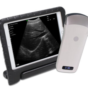
The VISION3 L is equipped with a dedicated linear probe and a streamlined hardware platform, ensuring efficient signal transmission and high-quality superficial tissue imaging. Its core hardware and probe parameters are systematically summarized in the following table, covering key indicators such as scanning mode, frequency, elements, and physical dimensions:
|
Technical Parameter
|
Detailed Specifications
|
|---|---|
|
Scan Mode
|
Electronic Array, Electronic Linear Array Scanning, ensuring uniform and high-resolution imaging for superficial tissues
|
|
Probe Type & Frequency
|
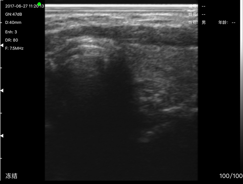
Linear Probe, dual-frequency adjustable: 7.5MHz/10MHz, optimized for superficial tissue observation
|
|
Elements
|
80 high-precision elements, enabling accurate capture of superficial tissue ultrasound signals with minimal distortion
|
|
Scanning Depth
|
40mm-100mm, continuously adjustable to adapt to different superficial and medium-depth tissue imaging needs
|
|
Field of View
|
Linear L40, providing a clear and focused imaging range for detailed observation of small organs and superficial structures
|
|
Image Gray Scale
|
256 levels, accurately reproducing subtle density differences of superficial tissues for precise lesion identification
|
|
Supported Display Screen
|
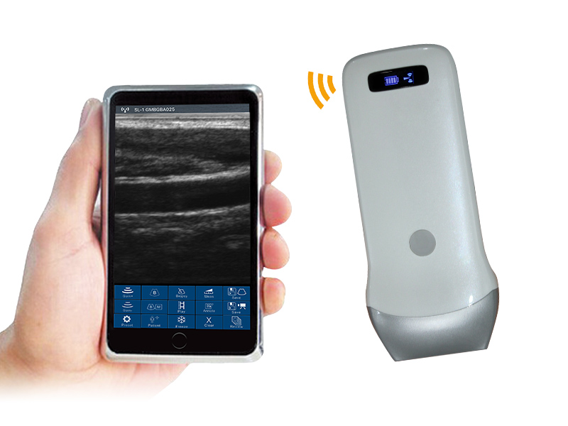
Adaptable to smartphone or tablet screens, enabling flexible and on-the-go imaging display
|
|
Supporting System
|
iOS, Android, Windows, realizing cross-system compatibility for diverse terminal matching
|
|
Size
|
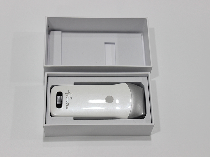
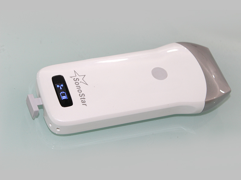
156mm×60mm×20mm, compact and ergonomic design for comfortable handheld operation
|
|
Net Weight
|
250 grams, lightweight structure for convenient portability and long-duration clinical use
|
|
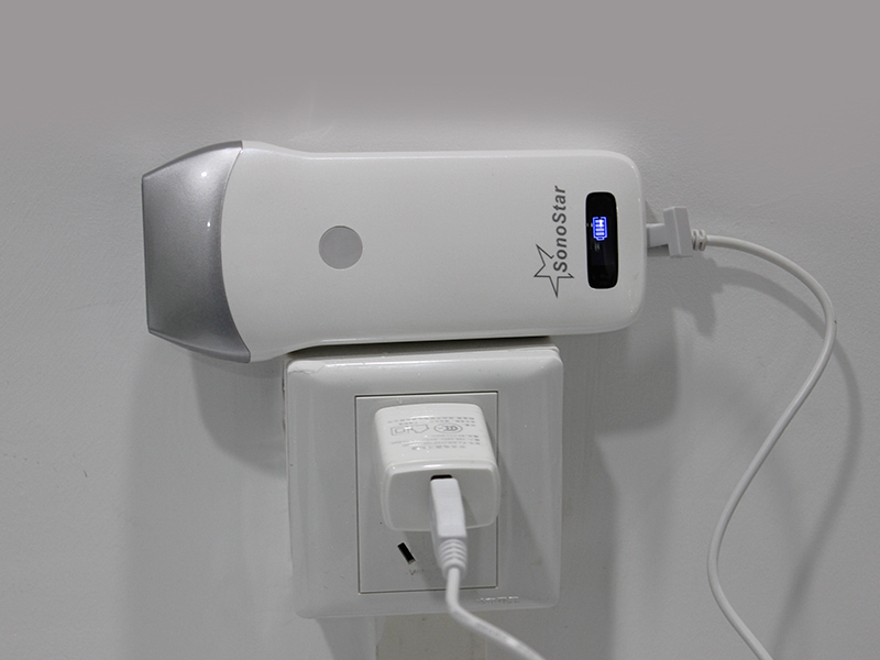
Battery Capacity
|
Built-in lithium battery, 4200mAh high capacity for stable power supply
|
|
Battery Working Time
|
3 hours of continuous operation, fully covering daily clinical examination needs without frequent charging
|
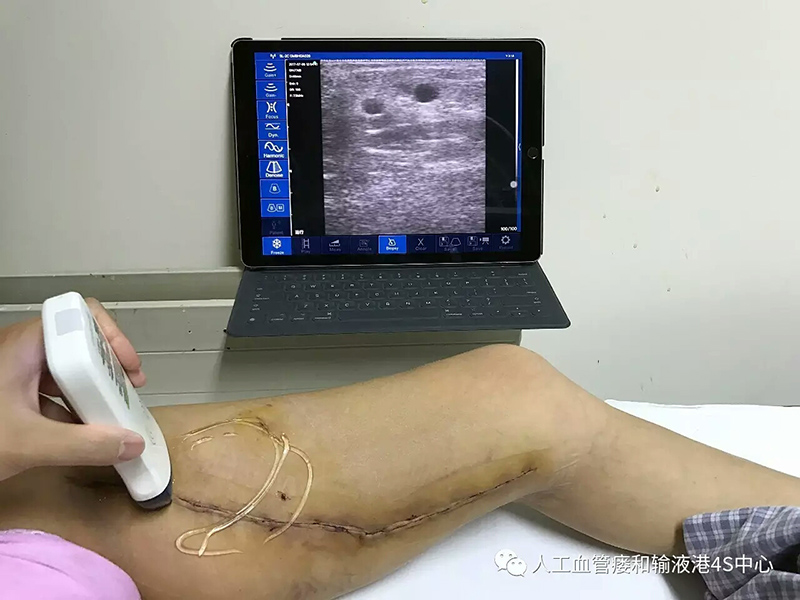
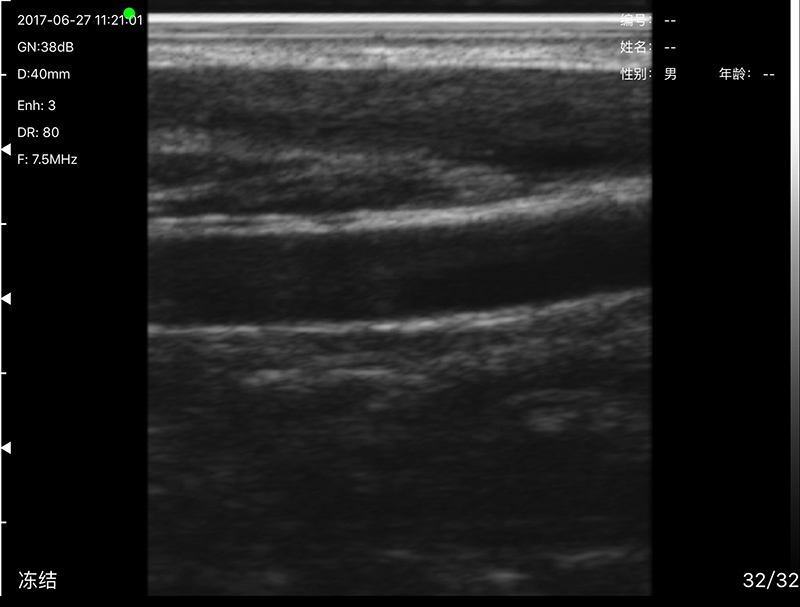
The 80-element configuration of the VISION3 L is tailored for linear array scanning, ensuring high signal-to-noise ratio and clear imaging for superficial tissues. The dual-frequency linear probe (7.5MHz/10MHz) offers strong adaptability: 7.5MHz is suitable for medium-superficial tissue imaging (e.g., musculoskeletal structures, thyroid glands) with balanced resolution and penetration; 10MHz high frequency delivers ultra-clear imaging for superficial small organs (e.g., breast nodules, superficial blood vessels), enabling the display of subtle structural details. With a scanning depth range of 40mm-100mm and L40 field of view, the system can accurately cover most superficial tissue examination scenarios, ensuring detailed observation of target areas.
The compact size (156mm×60mm×20mm) and 250-gram weight make the VISION3 L easy to carry in a medical bag, suitable for bedside examinations, home visits, and outdoor medical services. The 4200mAh high-capacity battery supports 3 hours of continuous work, eliminating power concerns during mobile clinical operations, while the compatibility with smartphones and tablets enhances its on-the-go usability.
2. Imaging Performance & Adjustment Functions
The VISION3 L delivers excellent imaging quality for superficial tissues, supported by targeted display modes and precise image adjustment functions, ensuring clear, stable, and detailed imaging effects to assist clinical diagnosis.
2.1 Display Modes
The system supports two core imaging display modes, optimized for superficial tissue observation and dynamic monitoring:
-
B-mode: Real-time two-dimensional gray-scale imaging, providing clear anatomical structure display of superficial tissues and small organs. It serves as the core mode for routine examinations, enabling doctors to directly observe tissue morphology and identify abnormal lesions.
-
B/M-mode: Combines B-mode two-dimensional imaging with M-mode time-axis display, suitable for observing dynamic changes of superficial moving tissues (e.g., blood vessel pulsation, muscle contraction). This mode is particularly useful for evaluating the functional status of superficial structures.
The 256-level image gray scale ensures that the VISION3 L can accurately reproduce subtle density differences between normal superficial tissues and lesions, such as small nodules, vascular wall thickening, and musculoskeletal tissue injuries, providing a clear basis for clinical diagnosis.
2.2 Image Adjustment Functions
To optimize imaging quality for different patients and examination sites, the VISION3 L is equipped with a set of precise image adjustment functions, supporting personalized imaging optimization:
-
Gain: Adjustable range of 30dB-105dB, allowing precise adjustment of image brightness. It can highlight tissue details in different depth layers of superficial structures, avoiding overexposure of shallow tissues or underexposure of medium-superficial tissues.
-
Noise Reduction: 5-level adjustable (0-1-2-3-4), with intensity increasing from level 0 to 4. It effectively filters out interference signals in superficial tissue imaging, reducing image noise and ensuring imaging stability and smoothness.
-
Dynamic Range: 8-level adjustable (40/50/60/70/80/90/100/110), adjusting the range of gray-scale levels displayed in the image. It balances the display of high-density and low-density superficial tissues, improving image层次感 and detail recognition.
These adjustment functions work together to ensure that the VISION3 L can obtain optimal imaging effects in various superficial tissue examination scenarios, from small organ inspections to musculoskeletal imaging, providing reliable visual support for accurate clinical diagnosis.
3. Clinical Auxiliary & Data Management Functions
The VISION3 L is equipped with practical clinical auxiliary functions and flexible data management capabilities, improving the efficiency of clinical examinations and facilitating data archiving and sharing.
3.1 Measurement Functions
The system integrates rich clinical measurement functions, covering routine and specialized measurement needs for superficial tissues and small organs:
-
Basic Measurement: Distance (length, thickness) and area measurement, suitable for evaluating the size of superficial lesions (e.g., thyroid nodules, breast masses), the thickness of organ walls, and the scope of abnormal tissues.
-
Obstetric Measurement: Supports routine prenatal examination parameters related to superficial fetal structures, such as fetal facial features, limbs, and superficial soft tissues, providing auxiliary data support for prenatal evaluation.
-
Other Measurements: Adapts to clinical needs such as angle measurement (for musculoskeletal joint examinations) and small organ volume calculation, meeting diverse diagnostic requirements for superficial tissue imaging.
All measurement results comply with international medical measurement standards, with high data accuracy and repeatability, ensuring that clinical diagnosis is supported by reliable quantitative data.
3.2 Data Storage Functions
The VISION3 L features flexible data storage capabilities, designed to facilitate clinical data management and on-the-go access:
Images and videos captured during examinations can be directly stored in smartphones or tablet PCs, eliminating the need for additional storage equipment. This design not only simplifies data storage operations but also enables convenient on-site review of examination results and quick sharing with colleagues for case discussion. The storage method is compatible with the file management systems of iOS, Android, and Windows devices, ensuring that data can be easily archived, exported, or uploaded to medical record systems, meeting clinical data management requirements.
4. System Compatibility & Portability Advantages
The VISION3 L emphasizes system compatibility and portability, adapting to diverse clinical scenarios and terminal devices, and improving the flexibility and efficiency of medical work.
4.1 System Compatibility
The system supports three major operating systems: iOS, Android, and Windows, and can be seamlessly connected to smartphones, tablets, and other terminal devices. This cross-system compatibility allows medical professionals to use their familiar devices for operation, avoiding the learning cost of new terminals and improving work efficiency. The device can quickly synchronize imaging data and operation commands with terminal devices, ensuring stable and smooth operation during examinations.
4.2 Portability Advantages
With a net weight of 250 grams and a compact size of 156mm×60mm×20mm, the VISION3 L is extremely portable and can be easily carried in a pocket or medical bag. It is suitable for a variety of mobile clinical scenarios, including community health services, home visits for the elderly and infirm, outdoor emergency rescues, and on-site examinations in remote areas. The 4200mAh high-capacity battery supports 3 hours of continuous operation, which is sufficient for daily clinical examinations. Its lightweight and portable design also reduces the burden of long-time handheld operation for medical professionals, improving work comfort.
5. Summary
The VISION3 L linear probe ultrasound system is a high-performance portable medical imaging device tailored for superficial tissue imaging. It integrates a dedicated linear probe, stable imaging performance, and practical clinical functions, with the core advantages of precision, portability, and user-friendliness. The 80-element hardware configuration, dual-frequency linear probe (7.5MHz/10MHz), and adjustable imaging parameters ensure high-quality imaging for superficial tissues and small organs, providing clear visual support for clinical diagnosis.
The system’s rich measurement functions, flexible data storage, and cross-system compatibility improve clinical work efficiency, while its compact and lightweight design enhances portability, adapting to diverse mobile clinical scenarios. Whether in routine small organ examinations, musculoskeletal evaluations, or primary healthcare services, the VISION3 L can provide reliable and professional imaging solutions. It is a practical and cost-effective portable ultrasound device, helping to improve the level of clinical diagnosis and service efficiency in various medical settings.
Your message has been sent

Reviews
There are no reviews yet.