
Description
1. Core System Overview & General Functions
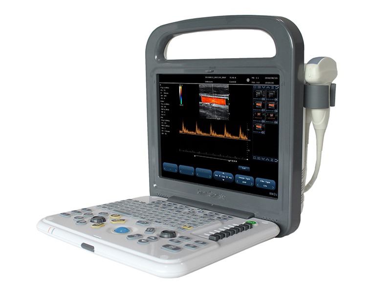
The VISION C8 is built on a PC platform, ensuring stability, expandability, and seamless integration with modern healthcare workflows:
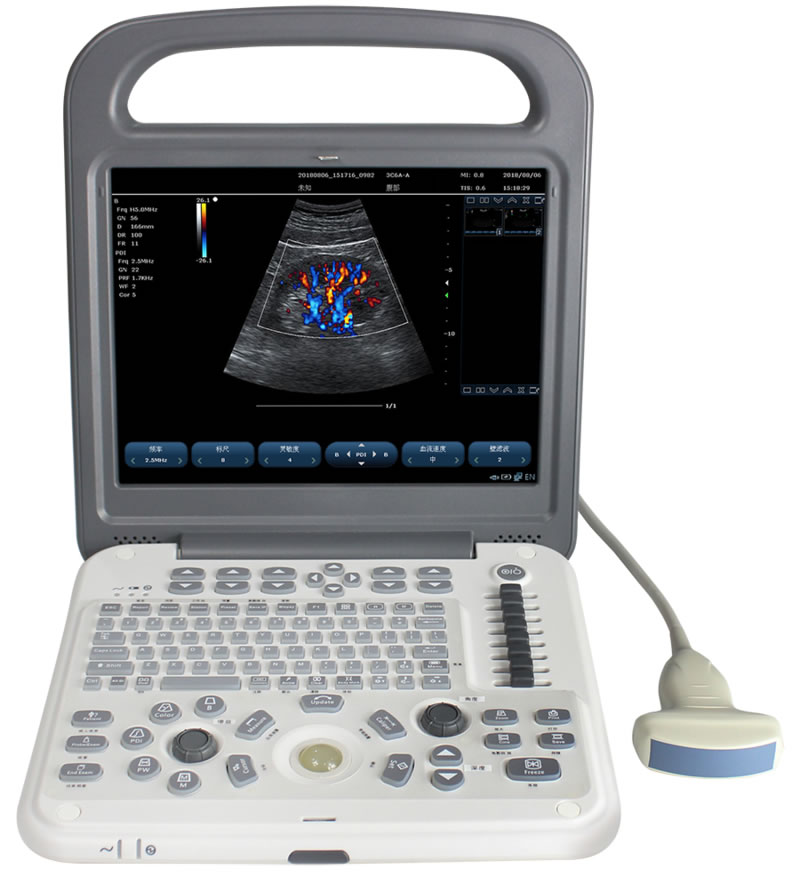
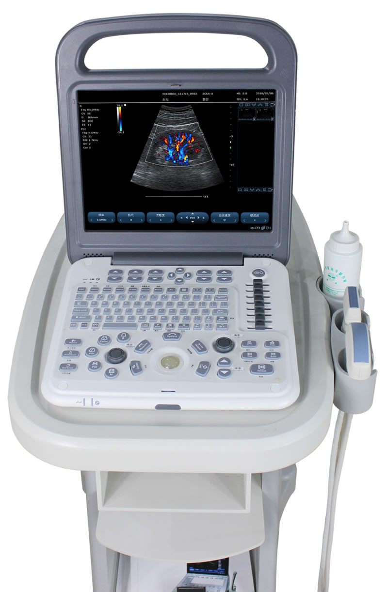
- Display & Ergonomics: 12.1-inch high-resolution color LCD monitor provides bright, sharp image visualization with wide viewing angles, supporting accurate diagnosis even in varied lighting conditions. The system weighs only 4.5kg, enhancing portability for mobile use while maintaining robust performance.
- Power Supply: Dual power system with built-in large-capacity lithium battery (2 hours of continuous operation) and AC power support. The screen displays real-time battery status, ensuring uninterrupted use during field visits or power outages.
- Quick Startup & Operation: Cold start in just 39 seconds enables rapid deployment, critical for emergency scenarios or high-volume clinics. Main interface miniatures simplify navigation, while full-screen Chinese/English annotation input (including two Chinese input methods, such as Wubi) caters to diverse user needs.
- Patient Data Management: Built-in patient data management station and customized comment functions (insert, edit, save) streamline documentation. A 500G hard disk provides ample storage for static images, dynamic videos, and medical records, supporting comprehensive archiving and playback.
2. Advanced Imaging Technologies & Modes
The VISION C8 integrates cutting-edge imaging technologies to optimize clarity, contrast, and diagnostic accuracy:
-
Key Imaging Functions:
- Color Doppler Enhancement Technology: Improves blood flow visualization, enhancing detection of vascular abnormalities.
- PHI Pulse Inverse Phase Tissue Harmonic Imaging + Frequency Composite Technique: Minimizes artifacts and enhances tissue contrast, particularly useful for deep or obese patients.
- Spatial Composite Imaging: Reduces speckle noise and improves spatial resolution by combining multiple scan angles.
- Linear Array Probe Independent Deflection & Linear Trapezoidal Spread Imaging: Expands the field of view for linear probes, reducing probe repositioning during superficial tissue examinations.
- B/Color/PW Trisynchronous Technology: Simultaneously displays 2D imaging, color Doppler, and pulsed wave Doppler for real-time structural and functional assessment.
- Multibeam Parallel Processing: Accelerates image acquisition and processing, ensuring smooth real-time imaging (up to 80 frames per second).
- Speckle Noise Suppression & B-Mode Enhancement: Refines image texture and reduces background interference, improving the visibility of subtle lesions.
-
Imaging Modes: Supports a comprehensive range of modes to adapt to diverse clinical needs:
- Foundational Modes: B Mode (2D), 2B Mode (dual B-mode), 4B Mode (quad B-mode), M Mode (motion tracking).
- Doppler Modes: Color Doppler, Power Doppler Imaging (PDI), Pulsed Wave (PW) Doppler, Continuous Wave (CW) Doppler.
- Combined Modes: B+C+PW (trisynchronous), B+PDI+PW (trisynchronous), Dissection M Mode.
- Display Options: Single-window, dual-window real-time, or four-window display for multi-view analysis.
3. Probe Specifications & Compatibility
The VISION C8 offers flexible probe options to cover all major clinical applications, with advanced frequency tuning for optimal imaging:
- Probe Interface: Zero-force metal body connector with two mutually compatible interfaces, supporting easy probe swapping without damaging connectors.
- Frequency Range: 2.0–10MHz variable frequency, with each probe offering 5 adjustable frequencies (fundamental and harmonic) to adapt to different tissue depths and imaging needs.
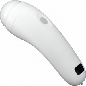
- Optional Probes:
- Convex Probe: For abdominal, pelvic, and obstetric imaging (2.5–6.0MHz).
- Linear Probe: For superficial tissues (e.g., thyroid, skin, musculoskeletal) (5.0–10MHz).
- Transvaginal Probe: For gynecological and early obstetric examinations (5.0–9MHz).
- Phased Array Probe: For cardiac imaging (2.0–3.5MHz).
- Microconvex Probe: For pediatric or small-organ imaging.
- Special Features: Optional puncture guidance with adjustable puncture lines and angles, supporting precise interventional procedures (e.g., biopsy, vascular access). All probes are B/D dual-purpose (B-mode + PW Doppler), enabling simultaneous structural and hemodynamic assessment.
4. Two-Dimensional Grayscale Imaging Parameters
The VISION C8’s grayscale imaging capabilities ensure detailed visualization of anatomical structures:
- Imaging Performance:
- Scanning Depth: Up to ≥351mm, covering deep abdominal organs and superficial tissues.
- Imaging Speed: Maximum 80 frames per second, ensuring smooth visualization of dynamic processes (e.g., fetal movement, cardiac motion).
- Beam Focus: 4 adjustable focal points, allowing operators to sharpen clarity on specific target areas.
- Amplification: Unlimited real-time amplification and 3x freeze amplification for detailed analysis of small structures.
- Image Optimization:
- 8-segment TGC (Time-Gain Compensation) adjustment: Equalizes image brightness across different depths.
- Continuously adjustable 2D gain and visual acoustic power control: Fine-tunes image quality based on tissue properties.
- Digital Sound Beamformer: Supports full-range dynamic focusing, dynamic variable aperture, and dynamic trace, with focus positions adjustable across the entire imaging area.
- False Color Options: 7 preset false color maps for enhanced tissue differentiation.
- Preset Conditions: Organ-specific optimal imaging settings reduce operational adjustments, improving workflow efficiency.
5. Doppler Imaging & Blood Flow Assessment
Comprehensive Doppler capabilities enable accurate hemodynamic evaluation:
- Color Doppler Specifications:
- Continuously adjustable Doppler gain and color enhancement: Optimizes blood flow visualization for low- or high-velocity vessels.
- B+COLOR dual-screen display: Simultaneously shows grayscale anatomy and color flow, improving spatial correlation.
- Baseline adjustment (±15 grades): Reduces aliasing and enhances detection of reverse flow.
- Spectral Doppler (PW/CW) Parameters:
- PW Testing Range: 0–7.5m/s, supporting measurement of both low- and high-velocity blood flow (e.g., arterial stenosis).
- Maximum Measured Velocity: 7.5m/s (positive or reverse flow), enabling accurate assessment of hemodynamic abnormalities.
- Sampling Volume: 1–8mm adjustable (8+ levels), with precise positioning for targeted vessel analysis.
- Scaleplate (≥16 grades) and PRF (0.7kHz–9.3kHz adjustable): Adapts to diverse flow velocities.
- Automatic Envelope Measurement & Real-Time Calculation: Streamlines quantitative analysis of spectral data, reducing manual error.
6. Measurement & Analysis Tools
The VISION C8 offers a comprehensive suite of measurement functions for quantitative diagnostic support:
- General Measurements: Distance, area, circumference, volume, area ratio, distance ratio, angle, S/D velocity, time, heart rate, acceleration, and more—covering basic anatomical and hemodynamic assessments.
- Obstetric Measurements: Supports data collection for ≥3 fetuses, including fetal weight calculation, growth curve display, and fetal echocardiography (left ventricular function, myocardial weight). Three dedicated fetal measurement packages (OB1, OB2, OB3) cater to different gestational stages.
- Specialized Measurements:
- Blood flow sampling volume with 8+ adjustable levels.
- Automatic measurement of endovascular media, aiding in vascular disease staging.
- TEI Index calculation for cardiac function assessment.
- Flexible Data Display: All measurement data windows are removable, allowing customization of the imaging interface for unobstructed visualization.
7. Connectivity & Data Output
The VISION C8 features rich interfaces for seamless integration with healthcare systems and peripherals:
- Input/Output Interfaces: Digital signal input, VGA, S-Video, USB, audio, and network interfaces. Supports connection to printers (DeskJet, LaserJet, video printers), external monitors, and storage devices.
- DICOM Compatibility: DICOM3.0 interface enables integration with hospital PACS systems, facilitating remote consultation and centralized data management.
- Network Real-Time Transmission: User data can be transmitted to servers in real time, supporting telemedicine and collaborative diagnostics.
- Foot Switch Interface: Enables hands-free operation (e.g., image capture, freeze), enhancing efficiency during interventional procedures.
8. Clinical Applications & Target Scenarios
The VISION C8’s versatility makes it suitable for a wide range of clinical specialties:
- Primary Applications: Abdominal (liver, kidney, spleen), cardiac (structural and functional assessment), obstetrics/gynecology (fetal monitoring, pelvic imaging), vascular (carotid, peripheral vasculature), and superficial tissue (thyroid, musculoskeletal) examinations.
- Target Environments: Multi-specialty clinics, hospitals, mobile medical services, rural healthcare facilities, and emergency response teams. Its portability, long battery life, and quick startup make it particularly valuable for field-based diagnostics and remote healthcare delivery.
9. Conclusion
The VISION C8 Color Doppler Ultrasound Scanner combines advanced imaging technology, versatile functionality, and user-centric design to deliver reliable diagnostic performance. Its comprehensive imaging modes, flexible probe compatibility, and robust data management capabilities address the diverse needs of modern healthcare, while its portability and dual power supply expand its utility beyond traditional clinical settings. Whether used for routine screenings, complex diagnostic evaluations, or interventional guidance, the VISION C8 empowers healthcare professionals to deliver accurate, efficient care across multiple specialties. With its balance of performance, portability, and ease of use, it stands as a valuable tool for both established clinics and mobile medical services.

Reviews
There are no reviews yet.