

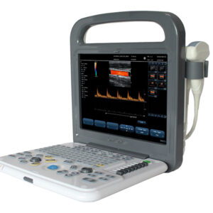
Description
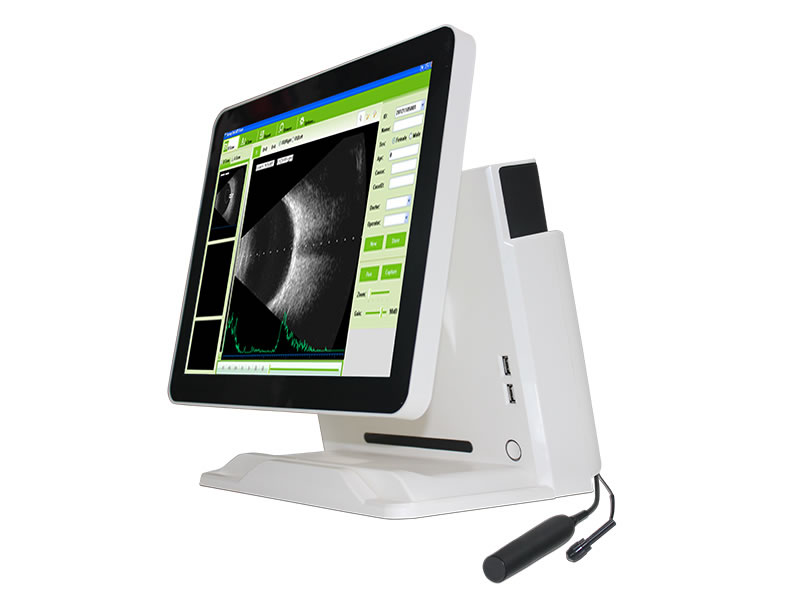
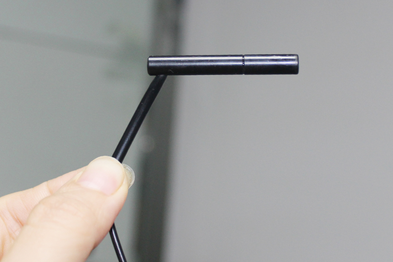
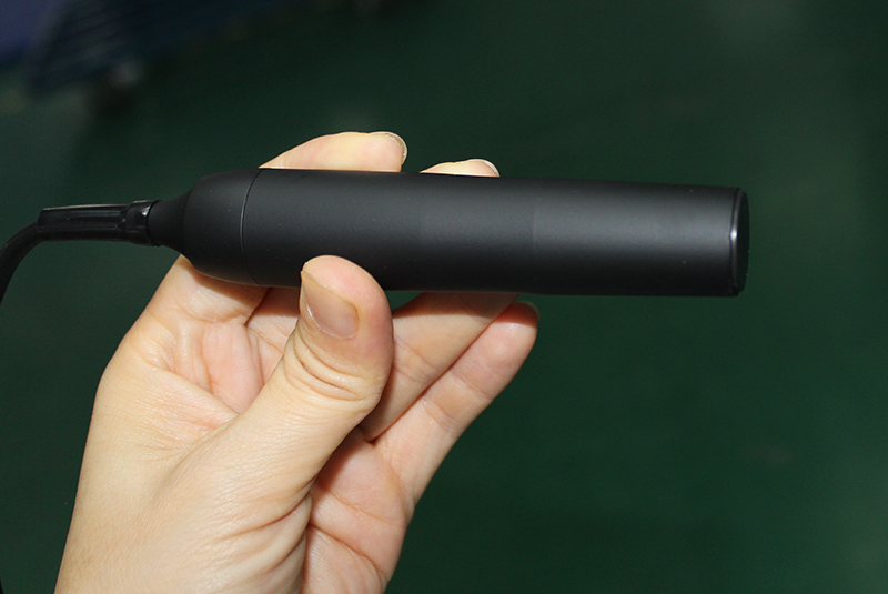
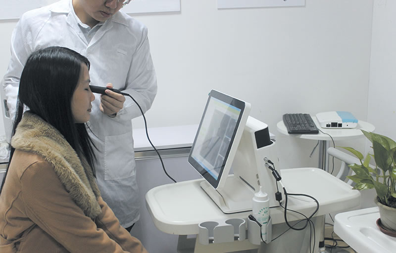
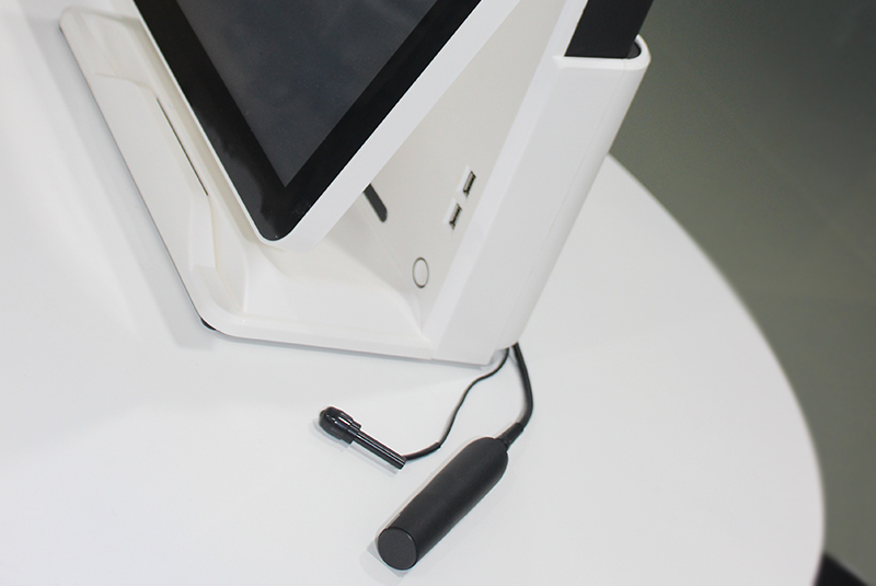
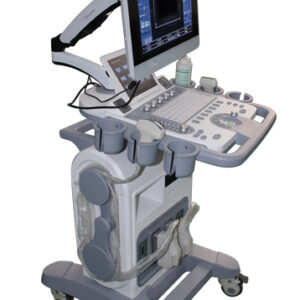
The SAB-500 Ophthalmic A/B Scanner is a high-precision diagnostic device tailored for comprehensive intraocular examination, designed to visualize and assess ocular structures with exceptional clarity. Equipped with advanced A-scan and B-scan modules, alongside specialized modes for vitreous body enhancement and retinal observation, it serves as an indispensable tool for ophthalmologists in diagnosing a wide range of intraocular diseases. This scanner accurately identifies the location, shape, and scope of lesions, as well as their anatomical relationship with surrounding tissues, enabling precise diagnosis of conditions such as vitreous opacity, retinal detachment, and fundus tumors. Additionally, the A-scan function provides precise biometric measurements for intraocular lens (IOL) power calculation, supporting cataract surgery planning. Below is a detailed breakdown of its technical specifications, organized by functional modules to highlight its diagnostic capabilities and clinical utility.
1. Core Clinical Functions & Diagnostic Scope
The SAB-500 integrates multi-mode imaging and measurement capabilities to address diverse ophthalmic diagnostic needs, with a focus on intraocular structure assessment and surgical planning:
-
Imaging Modes: Offers normal scanning mode, vitreous body enhancement mode, and retina observation mode. The enhanced modes optimize visualization of delicate intraocular structures, overcoming challenges posed by opaque media (e.g., cataracts, vitreous hemorrhage) to clearly display the vitreous body, retina, choroid, and other deep ocular tissues.
-
Disease Diagnosis: Specialized in identifying intraocular pathologies, including vitreous opacity (floaters), retinal detachment (rhegmatogenous, tractional, exudative), fundus tumors (melanoma, hemangioma), macular holes, choroidal detachment, and intraocular foreign bodies. It precisely maps lesion boundaries and their proximity to critical structures (e.g., optic nerve, macula), aiding in staging and treatment planning.
-
Surgical Support: The A-scan module provides accurate ocular biometrics for IOL power calculation, a critical step in cataract surgery. It supports multiple IOL formulas to accommodate different patient conditions (phakic, aphakic, dense cataracts), ensuring optimal postoperative visual outcomes.
2. B-Scan Technical Specifications
The B-scan module employs high-frequency ultrasound and magnetic-driven technology to deliver high-resolution 2D imaging of intraocular structures, ideal for evaluating posterior segment pathology:
-
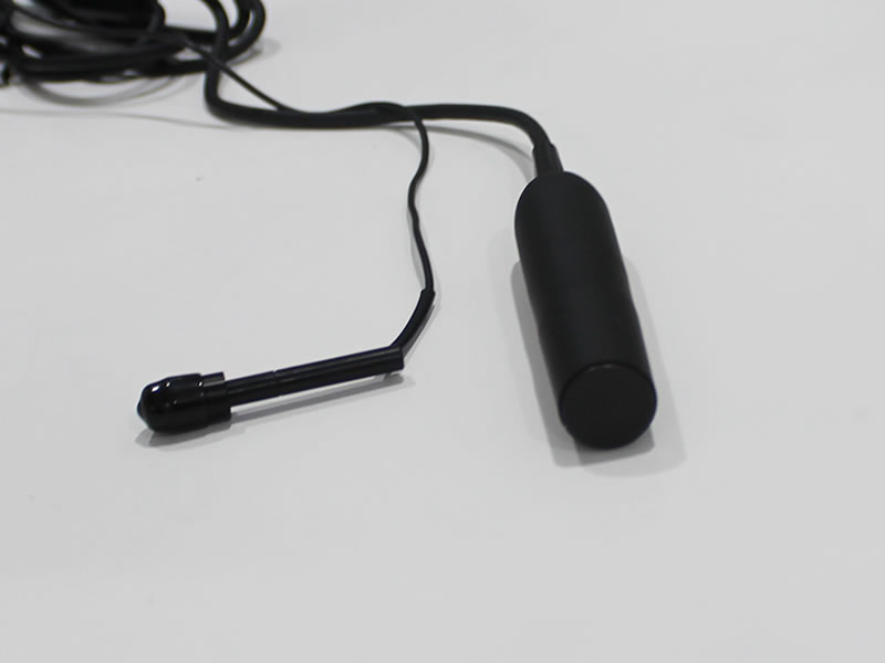
Frequency & Drive System: Optional dual-frequency configuration (10MHz/20MHz) to adapt to diverse diagnostic scenarios. The 10MHz frequency balances penetration and resolution for general posterior segment imaging, while the 20MHz frequency provides ultra-high resolution for detailed observation of superficial retinal layers and small lesions. Equipped with a magnetic-driven scanning mechanism, it operates noiselessly, ensuring patient comfort during examinations.
-
Scanning Parameters: Adopts sector scanning mode with a 53° scanning angle, covering a wide field of view of the posterior segment. Scanning depth reaches 60mm, sufficient to visualize the entire vitreous cavity, retina, choroid, and orbital tissues. Geometry position precision is exceptional: lateral ≤10% and vertical ≤5%, ensuring accurate localization of lesions.
-
Image Quality & Enhancement: Boasts superior resolution (lateral ≤0.3mm; vertical ≤0.2mm) to distinguish subtle anatomical details. 256-level grayscale imaging reproduces fine tissue density differences, while multi-color false color and OCT-like pseudo-color processing enhance contrast between pathological and normal tissues. Specialized vitreous body and retinal enhancement technologies highlight abnormal regions (e.g., vitreous opacities, retinal breaks) for easier identification.
-
Probe & Adjustment: Probe gain is continuously adjustable between 30dB-105dB, allowing operators to optimize signal strength based on patient ocular media clarity. Supports multi-continuous magnification and real-time magnification, enabling detailed observation of small lesions without compromising image quality.
-
Image Postprocessing & Storage: Equipped with multiple curve processing and pseudo-color curve processing tools to refine image details. Supports 100-image movie review for dynamic observation of intraocular structures, with image output in AVI (video) and JPG (static image) formats for documentation and consultation. Measurement functions include multi-group distance, perimeter, and area calculations to quantify lesion size and location.
3. A-Scan Technical Specifications
The A-scan module is a high-precision biometric tool for ocular axial length measurement and IOL power calculation, essential for cataract surgery planning:
-
Frequency & Display: 10MHz frequency with built-in LED indicator for real-time operation status. Scanning depth of 40mm covers the entire axial length of the eye, from the cornea to the posterior pole.
-
Measurement Precision & Parameters: Offers exceptional measurement precision (±0.05mm), ensuring reliable biometric data. Measures key ocular parameters: anterior chamber depth, lens thickness, vitreous body length, total axial length, and average values for bilateral comparison. Supports four eye modes to adapt to different clinical conditions: Phakic (with natural lens), Aphakic (without lens), Dense (opaque media, e.g., dense cataracts), and Various IOL (post-IOL implantation follow-up).
-
IOL Calculation & Statistics: Integrates six mainstream IOL formulas (SRK-II, SRK-T, HOFFER-Q, HOLLADAY, BINKHORST-II, HAIGIS) to calculate IOL diopter, accommodating diverse patient demographics (age, axial length, corneal curvature). Built-in statistical calculation provides average values and standard deviations for multiple measurements, reducing errors from single scans. Stores up to 10 scanning results per eye for comparative analysis and follow-up.
4. System Configuration & Operational Convenience
The SAB-500 is designed with user-centric features to streamline workflow and enhance diagnostic efficiency:
-
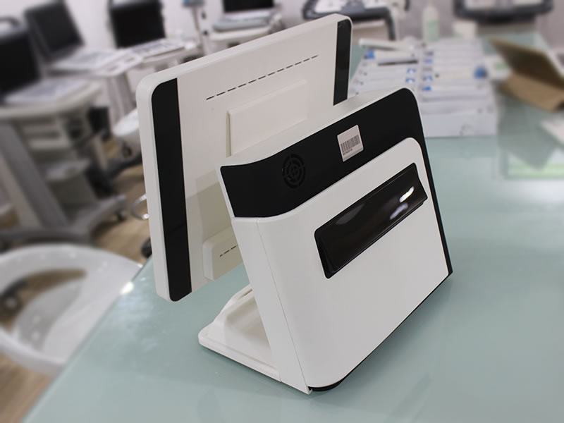
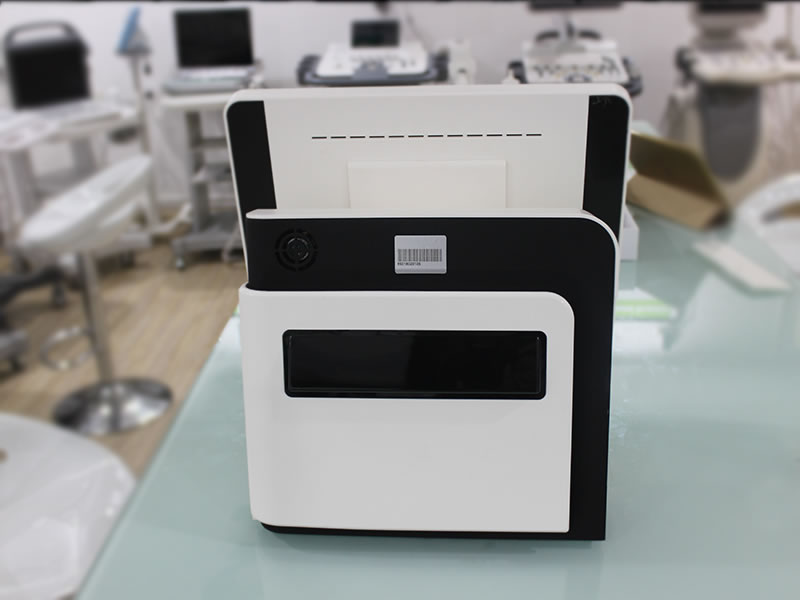
Display & Interface: Equipped with a 15-inch high-resolution LCD screen, providing clear, bright imaging with wide viewing angles for easy observation. Supports multiple display modes: single B-scan, dual B-scan (B+B), combined B+A scan, and single A-scan, adapting to different diagnostic needs.
-
Data Management: Features preset keywords for quick case labeling, multi-keyword case search for efficient retrieval of historical records, and user-defined report templates to customize documentation based on clinical requirements. This simplifies data management and enables seamless integration with electronic medical record (EMR) systems.
-
Power Supply: Built-in rechargeable battery offers up to 4 hours of continuous operation, supporting mobile examinations in clinics, operating rooms, or remote healthcare settings without relying on AC power. The battery status indicator provides real-time power information to avoid unexpected interruptions.
5. Clinical Applications & Target Users
The SAB-500 is widely applicable in ophthalmic clinics, hospitals, and surgical centers, catering to the following scenarios:
-
General Ophthalmology: Diagnosis of vitreoretinal diseases, intraocular tumors, and intraocular foreign bodies; evaluation of ocular trauma and choroidal detachment.
-
Cataract Surgery: Preoperative ocular biometrics and IOL power calculation; postoperative follow-up to assess IOL position and retinal status.
-
Retina Specialties: Detailed assessment of retinal detachment, macular pathologies, and vitreous-retinal interface disorders; monitoring of disease progression and treatment response.
-
Pediatric Ophthalmology: Examination of pediatric ocular structures (with gentle, noiseless operation) to diagnose congenital vitreoretinal abnormalities.
Target users include ophthalmologists, retinal specialists, cataract surgeons, and ophthalmic technicians seeking a reliable, high-precision tool for intraocular diagnosis and surgical planning.
6. Conclusion
The SAB-500 Ophthalmic A/B Scanner combines advanced imaging technology, precise measurement capabilities, and user-friendly design to deliver comprehensive intraocular diagnostic solutions. Its dual-frequency B-scan with specialized enhancement modes ensures clear visualization of delicate ocular structures, while the high-precision A-scan supports accurate IOL calculation for cataract surgery. With features such as noiseless operation, long battery life, and flexible data management, it adapts to diverse clinical environments, from routine clinics to mobile services. By enabling early and accurate diagnosis of intraocular diseases and optimizing surgical planning, the SAB-500 empowers ophthalmologists to improve patient outcomes and enhance clinical workflow efficiency, making it a valuable asset in modern ophthalmic practice.
Reviews
There are no reviews yet.