

Description
Sonostar Wireless Ultrasound Probe: Working Principle, Advantages, Specifications & Applications
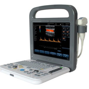
How It Works
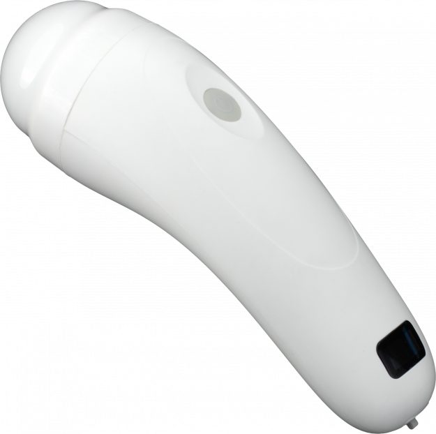
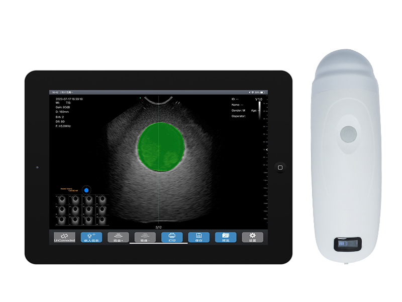
The Sonostar wireless ultrasound probe is a compact, screenless mini ultrasound scanner engineered for portability and clinical efficiency. We streamline the core components of a conventional ultrasound system into a miniaturized integrated circuit board housed directly within the probe. High-resolution ultrasound images are transmittedvia built-in private WiFi (no external WiFi or network connection required) to smartphones or tablets for real-time display. Visuals can be viewed simultaneously on both the connected smart device and tablet, enabling flexible clinical operation and seamless bedside monitoring.
Key Advantages
-
Ultra-Portable Design: All-in-one probe construction, compact and lightweight for effortless carrying, storage, and point-of-care use in any clinical setting.
-
Hassle-Free Wireless Connectivity: Stable one-tap WiFi pairing between the probe and tablet/phone, delivering intuitive, user-friendly operation with minimal setup time.
-
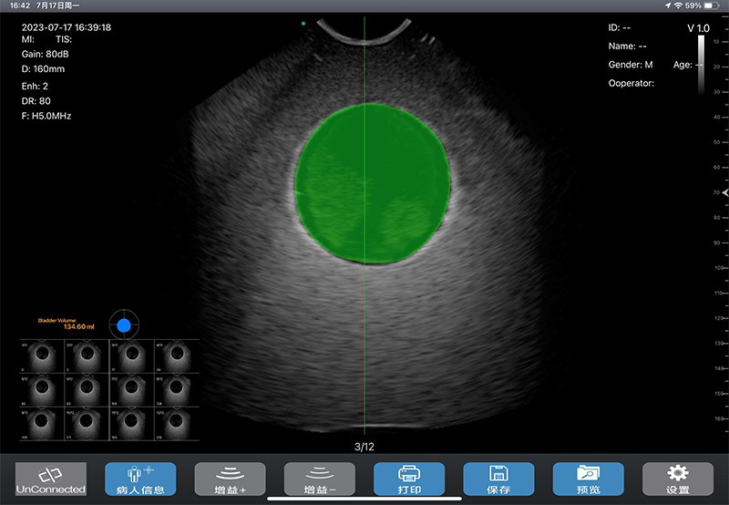
High Measurement Accuracy: Ultra-precise readings with a margin of errorless than 5%, ensuring reliable clinical data for diagnosis and decision-making.
-
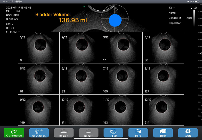
Fast Scanning Performance: Completed full scan and data processing in only 2 seconds, boosting clinical workflow efficiency and reducing patient waiting time.
-
Wide Measurement Range: Supports bladder volume measurement from 50ml to 2000ml, covering routine and critical clinical scenarios.
-
Premium 4D Electronic Convex Array Scanning: Equipped with high-channel-count, dense-array transducer configuration, producing crystal-clear imaging with zero mechanical scanning distortion for superior diagnostic clarity.
-
R20 Probe with Extended Coverage: Features an extra-wide scanning angle and expanded imaging coverage to capture full anatomical details effortlessly.
-
Maintenance-Free Precision Positioning: Built-in high-precision rotary encoder enables consistent, accurate positioning with no calibration required throughout the product lifecycle.
-
AI-Powered Advanced Measurement Algorithms: Integrated cutting-edge bladder wall recognition and precise edge detection technologies work in tandem with high-resolution transducers to maximize measurement accuracy. The proprietary algorithm is unaffected by bladder shape or size, with no restrictions based on patient selectivity, age group, or special conditions (e.g., post-hysterectomy bladder morphology). It delivers consistent, low-error readings even for small-volume or irregularly shaped bladders, with exceptional compatibility across all patient types. AI-enhanced recognition excels at identifying bladder walls with attached air bubbles and poorly defined edges; abnormal imaging datasets are integrated into model training to eliminate edge-hooking challenges and improve diagnostic reliability.
-
Real-Time Imaging for Clinical Guidance: Smooth, real-time scanning visuals support live visual monitoring during Foley catheter insertion and removal, preventing discomfort and complications caused by blind placement and enhancing patient safety.
Technical Specifications
-
Scan Mode: Electronic array probe, 4D scanning
-
Compatible Operating System: Apple iOS
-
Volume Measurement Range: 50ml – 2000ml
-
Automatic Measurement Error: < 5%
-
Scan & Processing Time: ≤ 2 seconds
-
Image Display Frame Rate: 15 frames/second
-
Case Data Storage: Stored via connected tablet
-
Printing: Compatible with external wireless thermal printer
-
Battery Performance: Built-in rechargeable battery; single full charge supports scanning for over 200 patients
-
Charging Method: USB cable charging
-
Product Dimensions: 170mm × 52mm × 55mm
-
Net Weight: 200g
Clinical Applications
-
Urology: Evaluate acute/chronic urinary retention severity; determine optimal catheterization and surgical timing; assess postoperative neuromodulation efficacy; diagnose urethral obstruction and benign prostatic hyperplasia (BPH) severity.
-
Obstetrics: Assess postpartum stress urinary incontinence; evaluate postpartum pelvic floor muscle injury degree.
-
Gynecology: Determine pelvic floor neuromuscular damage severity after pelvic tumor surgery; formulate targeted surgical or rehabilitation treatment plans.
-
Intensive Care Unit (ICU): Guide intermittent catheterization timing for long-term indwelling catheter patients; reduce catheterization frequency and infection risk; promote recovery of spontaneous urination.
-
Neurology: Evaluate detrusor and sphincter damage severity in patients with neurogenic bladder.
-
Nursing: Determine catheter removal timing for postoperative patients with indwelling catheters; shorten catheter indwelling duration and minimize healthcare-associated infection risk.
-
Rehabilitation Medicine: Monitor pelvic floor rehabilitation treatment efficacy; develop personalized bladder retraining and rehabilitation protocols; provide objective data for assessing bladder function recovery.
-
Emergency Department: Diagnose acute urinary retention and guide safe catheterization; assess bladder injury or rupture in acute trauma cases.
-
Radiotherapy: Monitor and evaluate bladder function in patients undergoing radiotherapy for pelvic floor tumors or bladder tumors.
Reviews
There are no reviews yet.