
Description
1. Core Scanning & Probe Parameters
Core hardware and probe specifications laying the foundation for deep-tissue imaging reliability:
-
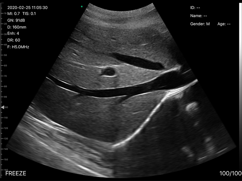
Scanning & Probe Configuration: Electronic Array & Electronic Convex Array Scanning, featuring a 128-element convex probe with dual-frequency adjustment (3.2MHz/5.0MHz). 3.2MHz ensures strong deep-tissue penetration (up to 305mm) for abdominal organs and mid-term pregnancy, while 5.0MHz balances resolution and penetration for medium-depth tissues. The multi-element design ensures uniform acoustic beam transmission and accurate capture of deep tissue signals.
-
Imaging Range: Adjustable convex scanning depth (90-305mm) to adapt to diverse deep-tissue thicknesses; Convex R60 field of view (60° arc) provides a wide imaging scope, facilitating comprehensive observation of abdominal, pelvic, and other deep anatomical structures without frequent probe movement.
-
Probe Physical Specs: The convex probe adopts an ergonomic curved-face design, ensuring close skin contact and uniform acoustic coupling. It matches the scanner’s 250g weight distribution, enabling comfortable handheld operation during prolonged deep-tissue examinations.
2. Imaging Quality & Optimization Functions
Comprehensive imaging modes and adjustment tools to enhance deep-tissue imaging clarity and diagnostic precision:
-
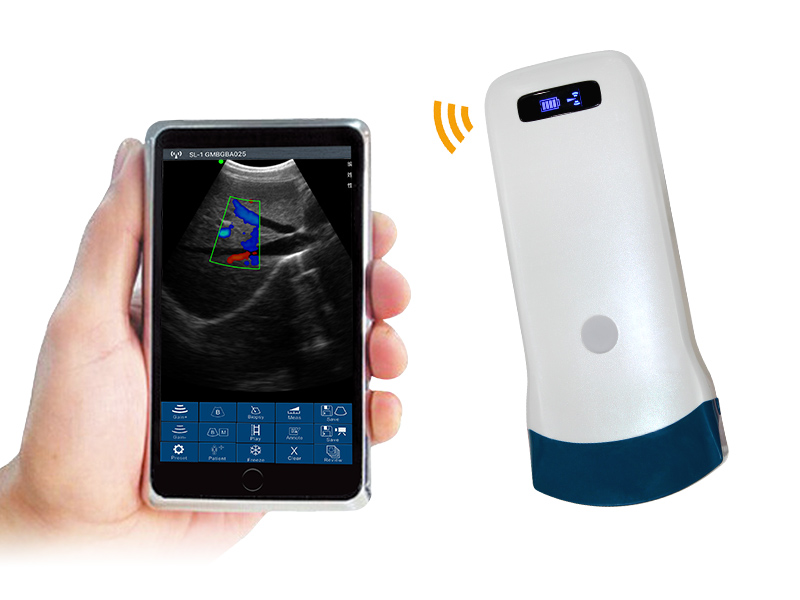
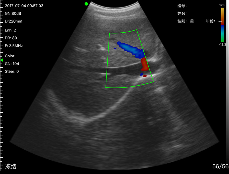
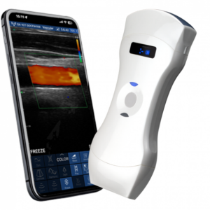
Imaging Modes: Supports B, B/M, Color, PDI (Power Doppler Imaging), and PW (Pulsed Wave Doppler) modes. It covers static deep-tissue morphology observation, dynamic process monitoring (e.g., fetal movement, vascular pulsation), and blood flow detection, providing multi-dimensional diagnostic information for deep tissues.
-
Imaging Foundation: 256-level image gray scale accurately reproduces subtle density differences between normal deep tissues and lesions, laying a solid foundation for precise identification of deep-seated abnormalities (e.g., abdominal tumors, pelvic cysts).
-
Image Adjustment: Equipped with comprehensive optimization tools, including Gain (30dB-105dB), 8-level Dynamic Range (40-110), and 5-level Noise Reduction (0-4). These tools effectively reduce deep-tissue imaging noise, balance brightness across different depths, and enhance image layering, further improving the clarity of deep-seated tissue structures.
3. Clinical Functions & Data Management
Practical clinical tools and flexible data handling to improve deep-tissue examination efficiency:
-
Measurement Functions: Supports distance, area, obstetrics, and other clinical measurements, providing accurate quantitative data. It assists in evaluating deep lesion size, organ dimensions, and obstetric indicators (e.g., fetal biparietal diameter), laying a reliable foundation for clinical decision-making and follow-up assessments.
-
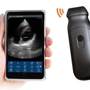
Data Storage: Images and videos are directly stored on mobile phones or tablets, eliminating the need for additional storage devices. This design facilitates on-site review of deep-tissue images, quick case sharing with colleagues, and easy data archiving for follow-up comparison and medical records.
4. System Compatibility & Physical Specifications
Cross-system compatibility and portable design optimized for mobile deep-tissue examination scenarios:
-
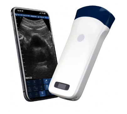
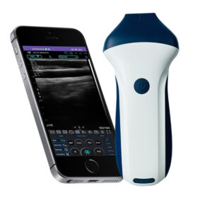
Display & Compatibility: Adapts to smartphone or tablet screens; compatible with iOS, Android, and Windows operating systems, enabling flexible connection to various terminal devices. This cross-system design reduces terminal replacement costs and adapts to different clinical usage habits of medical professionals.
-
Power Supply: Equipped with a built-in 4200mAh lithium battery, supporting 3 hours of continuous operation, fully covering daily clinical examination needs without frequent charging, ideal for mobile or bedside examinations.
-
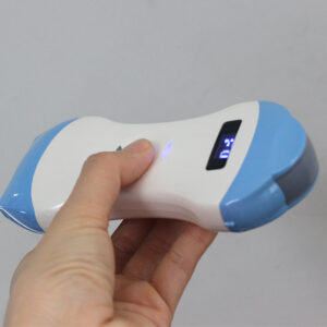
Physical Dimensions: 156mm×60mm×20mm compact size, 250g net weight, with an ergonomic handheld design for comfortable one-hand operation. It can be easily carried in a medical bag, suitable for bedside examinations, home visits, community healthcare, and field medical services requiring deep-tissue imaging.
The VISION5 C convex palm Doppler ultrasound scanner integrates 3.2/5.0MHz dual-frequency imaging, 128-element signal processing, and a 60° wide field of view. It provides stable and precise deep-tissue imaging solutions for diverse clinical scenarios, balancing deep penetration and diagnostic clarity. As a reliable tool for medical professionals, it meets the demands of routine deep-tissue examinations and specialized assessments in primary healthcare and clinical settings.
Your message has been sent

Reviews
There are no reviews yet.