
Description
1. Core Scanning & High-Frequency Probe Parameters
Foundational hardware that defines ultra-precise superficial imaging:
- Scanning & Probe Design: Electronic Array and Electronic linear array scanning, equipped with a high-frequency linear probe (16/20MHz) optimized for superficial tissue visualization. The dual-frequency configuration adapts to diverse clinical needs: 16MHz balances resolution and penetration for small organs (e.g., thyroid, mammary glands), while 20MHz delivers microscopic-level clarity for delicate structures (e.g., nerve bundles, superficial blood vessels, pediatric tissues).
- Imaging Range: Adjustable linear scanning depth (10-40mm) tailored to ultra-superficial to shallow-medium tissue examinations, covering the full spectrum of superficial anatomical structures. Linear L18 field of view (18mm) provides focused, high-density coverage, concentrating on target areas to eliminate background clutter and enhance the detection of small lesions or subtle abnormalities.
- Signal Precision: Advanced digital signal processing technology ensures high signal-to-noise ratio, minimizing image distortion and enhancing the definition of tissue boundaries—critical for distinguishing fine anatomical details (e.g., thyroid nodules, carotid plaques, musculoskeletal micro-tears).
2. Imaging Quality & Optimization Functions
Comprehensive tools to maximize clarity for high-frequency imaging:
- Imaging Modes & Foundation: Supports B, B/M, Color, PDI (Power Doppler Imaging), and PW (Pulsed Wave Doppler) modes. 256-level image gray scale accurately reproduces subtle tissue density differences, enabling the identification of early-stage lesions or structural variations that may be missed by lower-frequency scanners.
- Image Adjustment Suite: Equipped with multi-dimensional optimization features, including Gain (30dB-105dB), 8-level Dynamic Range (40-110), and 5-level Noise Reduction (0-4). These tools allow operators to fine-tune image brightness, contrast, and noise suppression, adapting to the unique properties of different superficial tissues (e.g., soft glandular tissue, dense muscle, delicate pediatric structures) for optimal diagnostic outcomes.
- Blood Flow Visualization: Enhanced Color and PDI modes provide clear visualization of micro-vascular networks, supporting accurate assessment of blood flow in superficial vessels (e.g., carotid arteries, mammary vasculature) and aiding in the diagnosis of vascular abnormalities.
3. Clinical Applications & Data Management
Specialized features tailored to diverse superficial imaging needs:
- Targeted Clinical Use Cases: Designed for a wide range of specialized applications, including thyroid nodule evaluation, small organ (parotid, submandibular) imaging, pediatric superficial structure assessment, vascular (carotid, peripheral) diagnosis, mammary gland examination, musculoskeletal micro-injury detection, and neurological (nerve bundle) visualization—meeting the demands of specialized clinicians and general practitioners alike.
- Measurement Capabilities: Supports precise quantitative measurements (distance, area, angle) for evaluating lesion size, tissue thickness, and vascular diameter. This data is critical for diagnosis, treatment planning, and long-term follow-up of conditions such as thyroid nodules, carotid stenosis, and musculoskeletal injuries.
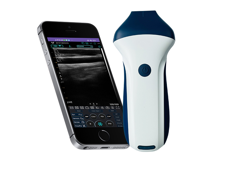
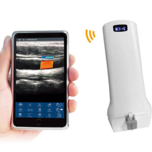
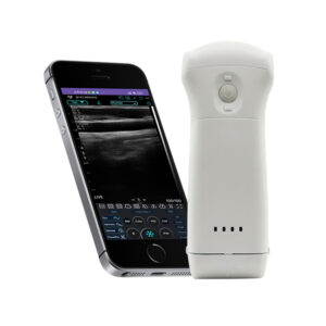
- Data Storage & Sharing: Images and videos are directly stored on mobile phones or tablets, eliminating the need for external storage devices. The wireless design facilitates on-site review, quick case sharing with colleagues, and seamless integration with electronic medical records (EMRs)—aligning with modern digital healthcare workflows.
4. Power & Physical Specifications
Portability and endurance optimized for specialized clinical use:
- Power Supply: Built-in 2200mAh lithium battery delivers 3 hours of continuous working time—ideal for daily clinical shifts, mobile visits, or specialized clinics with high patient volumes. The reliable battery performance ensures uninterrupted use during critical examinations.
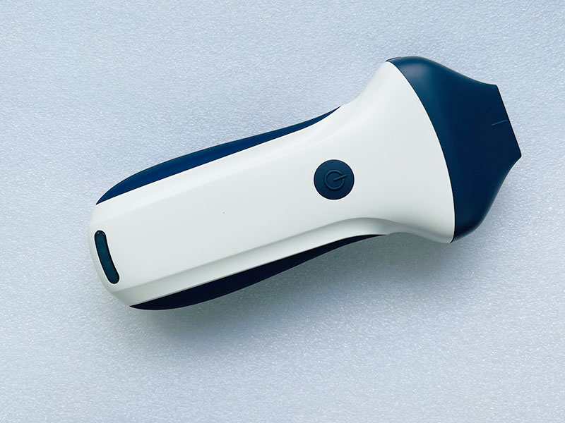
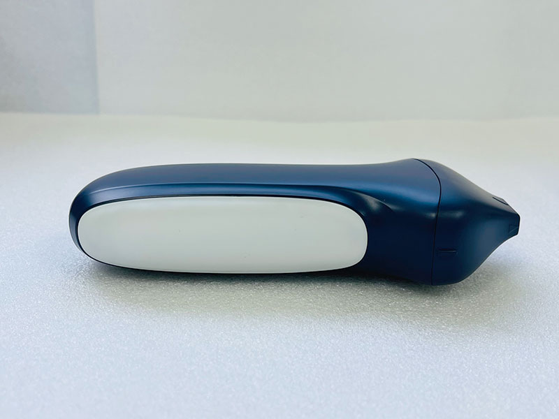
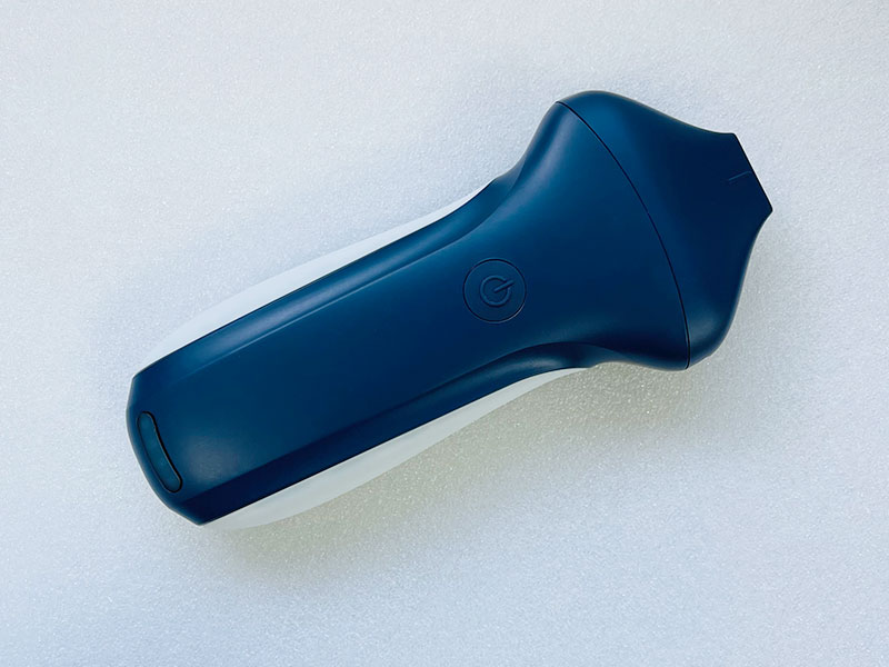
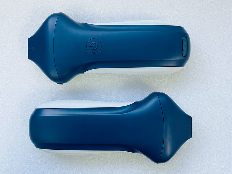
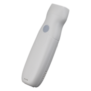
- Ultra-Portable Design: Compact dimensions (156mm×60mm×20mm) and 230g net weight make it easy to carry in a medical bag or pocket. Its lightweight, ergonomic form factor reduces operator fatigue during prolonged use, while the durable construction withstands the demands of daily clinical environments.
- System Compatibility: Seamlessly works with iOS, Android, and Windows operating systems, supporting flexible connection to smartphones, tablets, and computers. This cross-platform adaptability ensures compatibility with existing clinical setups and user preferences, enhancing its practicality in diverse healthcare settings.
The VISION7 H Linear Palm Doppler Ultrasound Scanner sets a new standard for high-frequency superficial imaging, combining microscopic resolution, specialized clinical versatility, and user-friendly portability. It is an indispensable tool for radiologists, endocrinologists, vascular surgeons, orthopedists, pediatricians, and general practitioners seeking precise, efficient imaging of superficial structures. Whether in a specialized clinic, hospital, or remote field setting, it delivers consistent, high-quality results to support timely and accurate clinical decisions, while its compact design ensures accessibility whenever and wherever specialized imaging is needed.

Reviews
There are no reviews yet.