
Description
1. Core Scanning & Probe Parameters
Core hardware and probe specifications laying the foundation for ultra-high-resolution superficial imaging:
-
Scanning & Probe Configuration: Electronic Array & Electronic Linear Array Scanning, equipped with a 128-element linear probe and dual ultra-high frequency adjustment (10MHz/14MHz). 10MHz balances resolution and penetration for medium-superficial tissues, while 14MHz delivers ultra-high resolution for tiny superficial structures (e.g., micro-nodules, thin blood vessel walls).
-
Imaging Range: Adjustable linear scanning depth (20-55mm) to adapt to diverse superficial tissue thicknesses; Linear L25 field of view provides a focused imaging scope, ensuring detailed observation of small target structures.
-
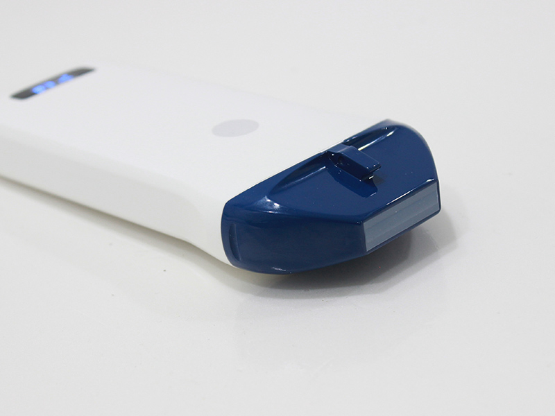
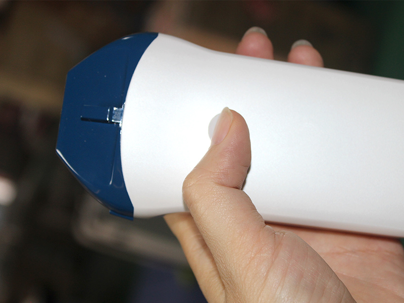
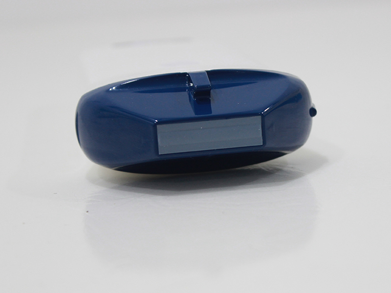
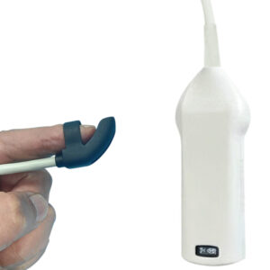
Probe Physical Specs: The linear probe features an ergonomic design, matching the scanner’s overall lightweight structure for comfortable handheld operation during prolonged examinations.
2. Imaging Quality & Optimization Functions
Comprehensive imaging modes and adjustment tools to enhance superficial imaging clarity and diagnostic precision:
-
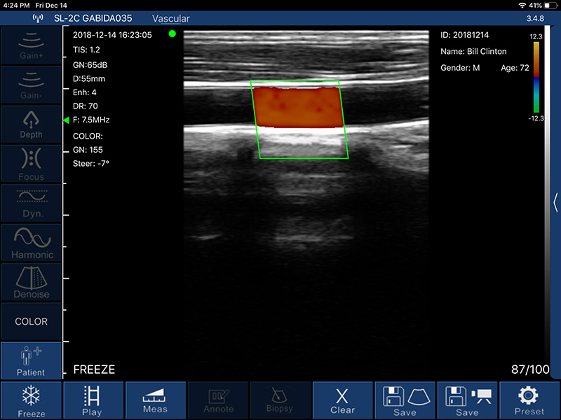
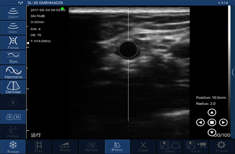
Imaging Modes: Supports B, B/M, Color, PDI (Power Doppler Imaging), and PW (Pulsed Wave Doppler) modes, covering static superficial tissue observation, dynamic process monitoring (e.g., vascular pulsation), and blood flow detection for multi-dimensional diagnosis.
-
Imaging Foundation: 256-level image gray scale accurately reproduces subtle density differences between normal tissues and lesions, laying a solid foundation for precise identification of tiny superficial abnormalities.
-
Image Adjustment: Equipped with multi-dimensional optimization tools, including Gain (30dB-105dB), 8-level Dynamic Range (40-110), and 5-level Noise Reduction (0-4). These tools effectively reduce image noise, balance tissue brightness, and enhance image layering, further improving the clarity of ultra-high-frequency imaging.
3. Clinical Functions & Data Management
Practical clinical tools and flexible data handling to improve superficial tissue examination efficiency:
-
Measurement Functions: Supports distance, area, obstetrics, and other clinical measurements, providing accurate quantitative data to assist in evaluating the size of tiny lesions, tissue thickness, and related physiological indicators.
-
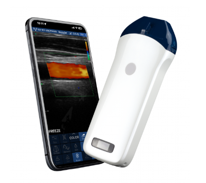
Data Storage: Images and videos are directly stored on mobile phones or tablets, eliminating the need for additional storage devices. This design facilitates on-site review of high-resolution images, quick case sharing with colleagues, and easy data archiving for follow-up comparison and medical records.
4. System Compatibility & Physical Specifications
Cross-system compatibility and portable design optimized for mobile clinical scenarios:
-
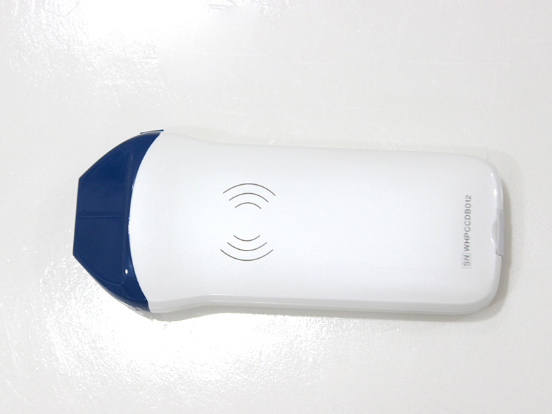
Display & Compatibility: Adapts to smartphone or tablet screens; compatible with iOS, Android, and Windows operating systems, enabling flexible connection to various terminal devices. This cross-system design reduces terminal replacement costs and adapts to different clinical usage habits of medical professionals.
-
Power Supply: Equipped with a built-in 4200mAh lithium battery, supporting 3 hours of continuous operation, fully covering daily clinical examination needs without frequent charging, ideal for mobile or bedside examinations.
-
Physical Dimensions: 156mm×60mm×20mm compact size, 200g net weight, ergonomic handheld design for comfortable one-hand operation. It can be easily carried in a medical bag, suitable for bedside, home visit, and field medical services.
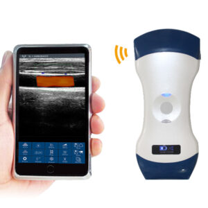
The VISION5 P UHF linear palm Doppler ultrasound scanner integrates 10/14MHz ultra-high-frequency imaging, 128-element signal processing, and ultra-portable design. It provides stable and precise ultra-high-resolution superficial tissue imaging solutions for diverse clinical scenarios, balancing diagnostic accuracy of tiny structures and operational flexibility, serving as a reliable tool for medical professionals in small organ and superficial tissue examinations.
Your message has been sent

Reviews
There are no reviews yet.