
Description
1. Core Hardware & Probe Parameters
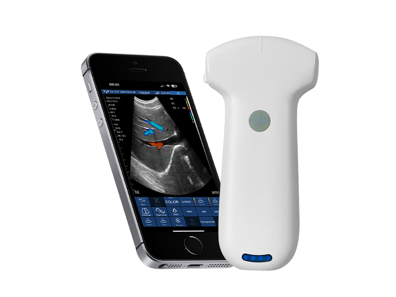
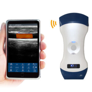
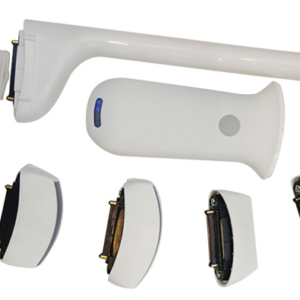
The VISION8 C is equipped with a high-performance hardware platform and a professional convex probe, laying a solid foundation for stable signal transmission and high-quality deep-tissue imaging. Its core hardware and probe parameters are summarized in the following table, covering key indicators such as elements, frequency, scanning range, and physical specifications:
|
Technical Parameter
|
Detailed Specifications
|
|---|---|
|
Scan Mode
|
Electronic Array Convex Scanning, ensuring stable and uniform imaging for deep tissues
|
|
Probe Type & Frequency
|
Convex Probe, dual-frequency adjustable: 3.2MHz/5.0MHz
|
|
Elements
|
192 high-density elements, enabling accurate acquisition of weak ultrasound signals
|
|
Channels
|
64 channels, supporting efficient signal transmission and optimizing imaging speed
|
|
Scanning Depth
|
90mm-305mm, continuously adjustable to adapt to different deep-tissue imaging needs
|
|
Field of View
|
Convex R60, providing a wide imaging range for comprehensive observation of abdominal and pelvic tissues
|
|
Image Gray Scale
|
256 levels, accurately reproducing tissue density differences for clear lesion identification
|
|
Size
|
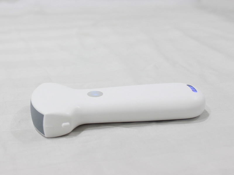
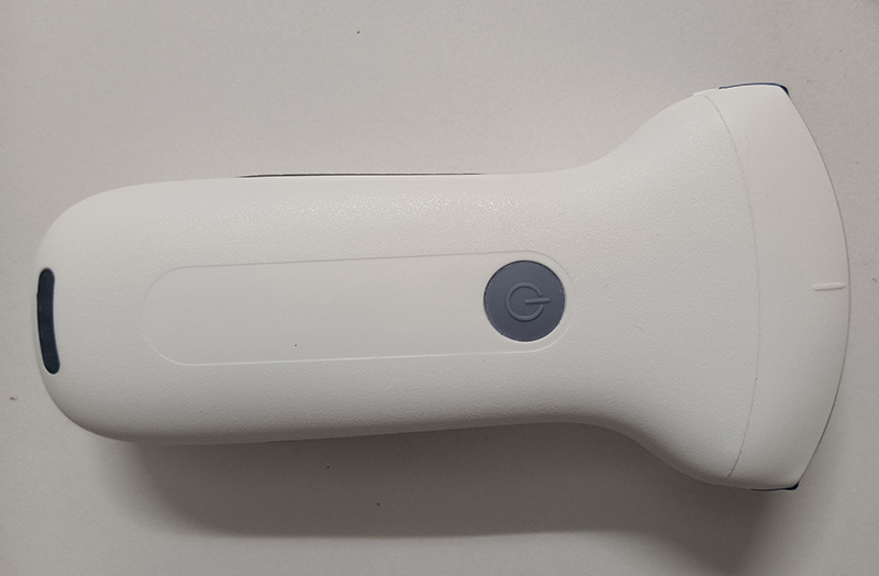
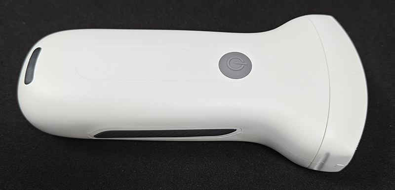
156mm×60mm×20mm, compact and ergonomic design for easy handheld operation
|
|
Net Weight
|
200 grams, ultra-lightweight for convenient portability and long-time use
|
|
Battery Capacity
|
Built-in lithium battery, 2200mAh (regular version); optional larger battery for extended use
|
|
Battery Working Time
|
Regular version: 1.5 hours; larger battery version: 3 hours of continuous operation
|
|
Charging Method
|
Wireless charging, supporting indefinite working time extension with continuous charging
|
|
WiFi Type
|
802.11n/2.4G/5G dual-band, maximum transmission rate 450Mbps
|
|
Supported Display Screen
|
Adaptable to iOS/Android/Windows screens (smartphones, tablets, computers)
|
|
Supporting System
|
iOS, Android, Windows (cross-system compatibility for flexible terminal matching)
|
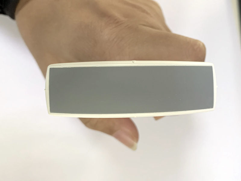
The 192-element and 64-channel configuration ensures that the VISION8 C can capture and process ultrasound signals with high efficiency, minimizing signal loss during transmission. The convex probe’s dual-frequency design (3.2MHz/5.0MHz) provides strong adaptability to different clinical scenarios: 3.2MHz is ideal for deep-tissue imaging (e.g., abdominal organs, late pregnancy fetus) with strong penetration, while 5.0MHz balances penetration and resolution for medium-depth tissue observation. With a scanning depth range of 90mm-305mm and R60 field of view, the system can cover most deep-tissue examination needs, ensuring comprehensive imaging of target areas.
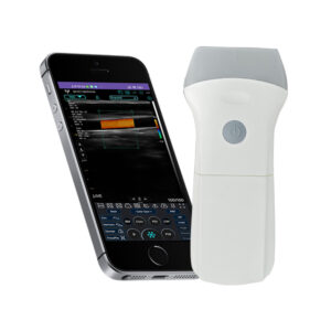
The ultra-lightweight design (200 grams) and compact size make the VISION8 C easy to carry and operate with one hand, suitable for bedside examinations, outdoor medical services, and remote primary healthcare. The 2200mAh battery and wireless charging function eliminate power constraints, supporting long-duration clinical work without relying on fixed power sources.
2. Imaging Performance & Display Functions
The VISION8 C delivers excellent imaging quality through a full range of display modes and precise image adjustment functions, ensuring clear, stable, and detailed imaging effects to support accurate clinical diagnosis.
2.1 Display Modes
The system supports five core imaging display modes, covering diverse clinical observation needs:
-
B-mode: Real-time two-dimensional gray-scale imaging, providing clear anatomical structure display of tissues and organs, serving as the basis for routine clinical examinations.
-
B/M-mode: Combines B-mode imaging with M-mode time-axis display, suitable for observing dynamic changes of moving tissues (e.g., fetal heart rate monitoring, abdominal organ peristalsis).
-
Color Imaging: Displays blood flow direction and distribution in color, helping to identify abnormal blood flow signals in abdominal organs, pelvic vessels, and other areas.
-
PDI (Power Doppler Imaging): Enhances weak blood flow signal display, suitable for detecting low-velocity blood flow in deep tissues or small vessels.
-
PW (Pulsed Wave Doppler): Enables accurate measurement of blood flow velocity, providing quantitative data support for diagnosing vascular diseases or evaluating fetal blood flow.
2.2 Image Adjustment Functions
To optimize imaging quality for different patients and examination sites, the VISION8 C is equipped with a comprehensive set of adjustable image parameters, supporting personalized optimization:
-
Gain: Adjustable range of 30dB-105dB, allowing precise adjustment of image brightness to highlight tissue details in different depth layers, avoiding overexposure or underexposure.
-
Focus: Multi-level focus adjustment, enabling concentration of ultrasound energy on the target area to improve imaging clarity of lesions or key anatomical structures.
-
Reverse Pulse Harmonics: Effectively reduces near-field noise and artifacts, improves image contrast, and enhances the display of subtle lesions that are easily obscured by noise.
-
Noise Reduction: 5-level adjustable (0-1-2-3-4), with intensity increasing from level 0 to 4, effectively filtering out interference signals to ensure image stability and smoothness.
-
Dynamic Range: 8-level adjustable (40/50/60/70/80/90/100/110), adjusting the range of gray-scale levels displayed in the image to balance the display of high-density and low-density tissues, improving overall image层次感.
With 256-level image gray scale, the VISION8 C can accurately reproduce the subtle density differences between normal tissues and lesions, enabling doctors to distinguish pathological changes more easily. The combination of multiple adjustment functions ensures that the system can obtain optimal imaging effects in various clinical scenarios, from deep abdominal examinations to medium-depth pelvic imaging.
3. Clinical Auxiliary Functions
The VISION8 C is equipped with a variety of clinical auxiliary functions, including precise measurement tools and professional puncture assistance, to improve the efficiency and accuracy of clinical diagnosis and interventional operations.
3.1 Measurement Functions
The system integrates rich clinical measurement functions, covering routine and specialized measurement needs:
-
Basic Measurement: Distance (length, thickness) and area measurement, suitable for evaluating the size of lesions, organ dimensions, and the scope of abnormal tissues.
-
Obstetric Measurement: Supports routine prenatal examination parameters, including fetal biparietal diameter, head circumference, abdominal circumference, femur length, and estimated fetal weight, providing accurate data support for fetal development evaluation.
-
Other Measurements: Adapts to clinical needs such as angle measurement and cyst volume calculation, meeting diverse diagnostic requirements.
All measurement results comply with international medical measurement standards, with high data accuracy and repeatability, ensuring reliable support for clinical diagnosis.
3.2 Puncture Auxiliary Function
For interventional clinical scenarios (e.g., abdominal puncture, pelvic cyst puncture), the VISION8 C is equipped with a professional puncture auxiliary system, including three core functions:
-
In-Plane Puncture Guide Line: The guide line is aligned with the imaging plane, enabling real-time observation of the puncture needle’s path to ensure accurate needle insertion into the target area.
-
Out-Plane Puncture Guide Line: Suitable for puncture operations requiring vertical needle insertion, providing clear guidance for needle entry point and depth.
-
Automatic Vascular Measurement Function: Quickly and accurately measures vascular diameter, blood flow velocity, and resistance index, providing a basis for vascular puncture and interventional treatment.
These functions significantly improve the accuracy, safety, and efficiency of puncture operations, expanding the clinical application scope of the VISION8 C to interventional diagnosis and treatment.
4. Data Storage & Transmission
The VISION8 C is equipped with a comprehensive data management system, supporting multiple formats of image and video storage, as well as high-speed wireless transmission, facilitating clinical data archiving, sharing, and remote consultation.
4.1 Storage Functions
The system supports multi-format storage of images and videos, including JPG, PNG, MP4, and DCM, adapting to different data usage needs:
-
JPG/PNG: Suitable for static image storage and quick sharing, convenient for case discussion and medical records filing.
-
MP4: Used for dynamic video storage, recording the entire examination process to facilitate subsequent review and analysis.
-
DCM: Complies with the Digital Imaging and Communications in Medicine (DICOM) standard, enabling seamless integration with hospital information systems (HIS) and picture archiving and communication systems (PACS) for standardized data management.
Data can be directly stored in mobile phones, tablets, or other terminal devices, eliminating the need for additional storage equipment and facilitating on-site data review and off-site transmission.
4.2 Playback & Transmission
The VISION8 C supports both manual and automatic playback modes, with adjustable playback frames (100/200/500/1000 frames), allowing doctors to review the examination process frame by frame and capture key imaging details. In terms of data transmission, the dual-band WiFi module (2.4G/5G) supports a maximum transmission rate of 450Mbps, enabling high-speed wireless transmission of images and videos. This realizes real-time data sharing between the device and computers, cloud servers, or remote consultation terminals, facilitating multi-departmental collaboration and telemedicine services.
5. System Compatibility & Portability Advantages
The VISION8 C features strong system compatibility and excellent portability, adapting to diverse clinical scenarios and terminal devices, and improving the flexibility of medical work.
5.1 System Compatibility
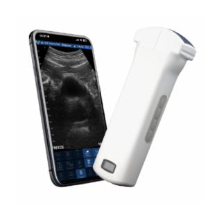
The system supports three major operating systems (iOS, Android, Windows) and can be connected to various terminal devices such as smartphones, tablets, and laptops. This cross-system compatibility eliminates the limitations of a single terminal, allowing medical professionals to use familiar devices for operation, improving work efficiency and user experience.
5.2 Portability Advantages
With a net weight of only 200 grams and a compact size of 156mm×60mm×20mm, the VISION8 C can be easily carried in a medical bag, suitable for mobile medical services such as home visits, outdoor free clinics, and emergency rescues. The 2200mAh battery (1.5-hour working time) and optional larger battery (3-hour working time) meet the needs of short-term and long-term clinical work, while wireless charging ensures continuous power supply, making the system completely free from power cord constraints.
6. Summary
The VISION8 C convex probe ultrasound system integrates advanced hardware configuration, excellent imaging performance, and rich clinical functions, with the core advantages of portability, high precision, and strong adaptability. Its 192-element and 64-channel hardware platform, combined with a dual-frequency convex probe, ensures high-quality deep-tissue imaging for abdominal, obstetric, and pelvic examinations. The comprehensive image adjustment functions and clinical auxiliary tools improve the accuracy and efficiency of diagnosis and interventional operations, while multi-format storage and high-speed wireless transmission facilitate data management and sharing.
With cross-system compatibility and ultra-lightweight design, the VISION8 C is applicable to a variety of clinical scenarios, including hospitals, clinics, primary healthcare institutions, and mobile medical services. It provides reliable and professional imaging solutions for medical professionals, helping to improve the level of clinical diagnosis and service efficiency, and is a highly practical portable convex probe ultrasound device.
Your message has been sent

Reviews
There are no reviews yet.