
Description
1. Core Scanning & Probe Parameters
Foundational hardware that ensures balanced imaging performance:
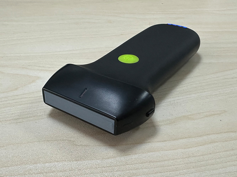
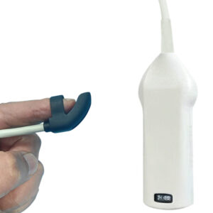
- Scanning & Probe Design: Electronic Array and Electronic linear array scanning, equipped with a 128-element linear probe for stable signal capture and processing. The dual-frequency configuration (7.5/10MHz) adapts to diverse needs: 7.5MHz balances resolution and penetration for medium-depth structures (e.g., musculoskeletal tissues, deeper small organs), while 10MHz delivers sharp visualization of superficial targets (e.g., thyroid nodules, superficial blood vessels, pediatric tissues).
- Imaging Range: Adjustable linear scanning depth (20-100mm) covers superficial to medium-depth tissue layers, eliminating the need for multiple devices for different examination requirements. Linear L40 field of view (40mm) provides expansive coverage, reducing probe repositioning and improving clinical efficiency—especially useful for large-area imaging of musculoskeletal structures or vascular networks.
- Signal Integrity: The 128-element probe ensures uniform acoustic beam distribution, minimizing image distortion and enhancing the definition of tissue boundaries. Advanced digital signal processing further optimizes signal-to-noise ratio, supporting the detection of subtle abnormalities and small lesions.
2. Imaging Quality & Optimization Functions
Comprehensive tools to maximize clarity across clinical scenarios:
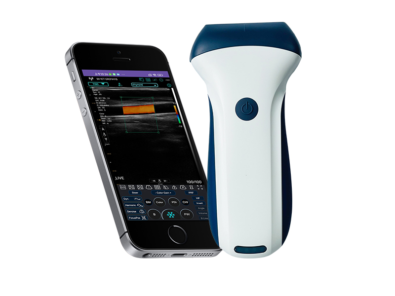
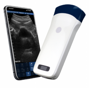
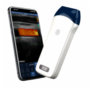
- Imaging Modes & Foundation: Supports B, B/M, Color, PDI (Power Doppler Imaging), and PW (Pulsed Wave Doppler) modes. 256-level image gray scale accurately reproduces subtle tissue density differences, enabling precise differentiation between healthy and abnormal tissues—critical for accurate diagnosis in diverse specialties.
- Image Adjustment Suite: Equipped with multi-dimensional optimization features, including Gain (30dB-105dB), 8-level Dynamic Range (40-110), and 5-level Noise Reduction (0-4). These tools allow operators to fine-tune image brightness, contrast, and noise suppression, adapting to tissue-specific properties (e.g., soft glandular tissue, dense muscle, fragile pediatric structures) for optimal visualization.
- Dynamic & Vascular Imaging: Enhanced Color and PDI modes provide clear real-time visualization of blood flow, supporting accurate assessment of vascular patency, perfusion, and velocity—key for diagnosing conditions like carotid stenosis, peripheral vascular disease, and abnormal vascularization in lesions.
3. Clinical Applications & Data Management
Practical features tailored to modern clinical workflows:
- Targeted Clinical Use Cases: Designed to excel in a broad range of applications, including thyroid nodule evaluation, small organ (parotid, submandibular) imaging, pediatric superficial structure assessment, vascular and carotid diagnosis, mammary gland examination, musculoskeletal injury detection (e.g., tendon tears, ligament sprains), and neurological (peripheral nerve) visualization. It meets the diverse needs of both specialized clinicians and general practitioners.
- Measurement & Storage: Supports precise quantitative measurements (distance, area, angle) for evaluating lesion size, tissue thickness, and vascular diameter—vital for diagnosis, treatment planning, and follow-up. Images and videos are directly stored on mobile phones or tablets, facilitating on-site review, quick case sharing, and seamless integration with electronic medical records (EMRs).
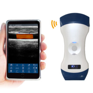
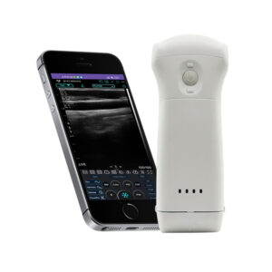
- User-Centric Operation: Compatible with smartphone or tablet screens, the scanner leverages intuitive touch-based controls via dedicated apps for iOS, Android, and Windows. This simplifies operation, reduces learning costs, and enables efficient use in fast-paced clinical environments.
4. Power & Physical Specifications
Portability and flexibility optimized for diverse use cases:
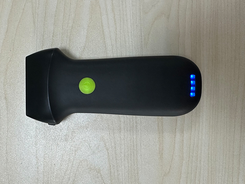
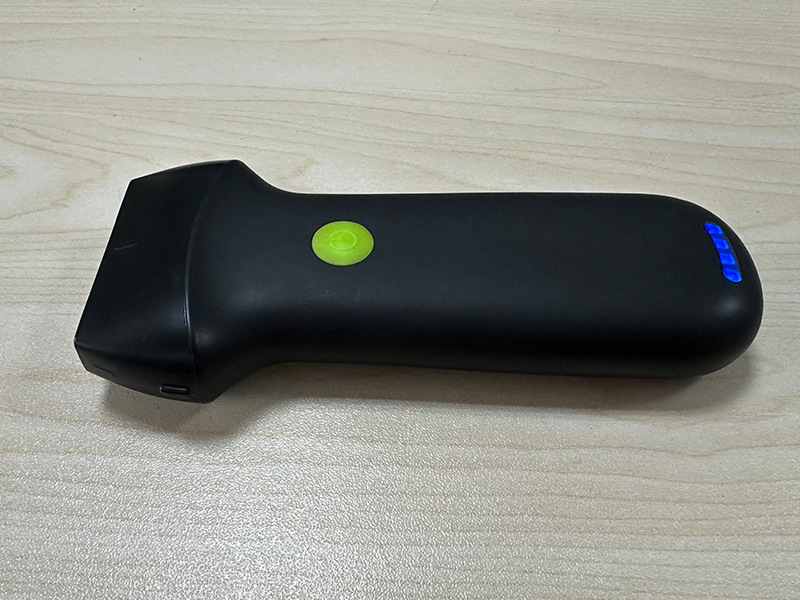
- Power Options: Built-in 2200mAh lithium battery with two versions—regular (1.5 hours of continuous use) for short shifts or quick examinations, and larger battery version (3 hours) for extended clinical work, mobile visits, or field services. This flexibility adapts to different practice needs without compromising performance.
- Ultra-Lightweight Design: Compact dimensions (156mm×60mm×20mm) and 180g net weight make it one of the lightest palm ultrasound scanners on the market. Its ergonomic form factor reduces operator fatigue during prolonged use, while durable construction withstands the demands of daily clinical and mobile environments.
- System Compatibility: Seamlessly works with iOS, Android, and Windows operating systems, supporting flexible connection to a wide range of devices. This cross-platform adaptability ensures compatibility with existing clinical setups and user preferences, enhancing its practicality across diverse healthcare settings.
The VISION7 L Linear Palm Doppler Ultrasound Scanner combines balanced imaging performance, versatile clinical applicability, and ultra-portability. It is an indispensable tool for radiologists, endocrinologists, vascular surgeons, orthopedists, pediatricians, and general practitioners seeking efficient, high-quality imaging of superficial to medium-depth tissues. Whether in a clinic, hospital, or remote field setting, it delivers consistent results to support timely and accurate clinical decisions, while its flexible design ensures accessibility and convenience for all users.

Reviews
There are no reviews yet.