

Description
1. Core Scanning & Probe Parameters
Core hardware and probe specifications laying the foundation for ultra-high-resolution superficial imaging:
-
Scanning & Probe Configuration: Electronic Array & Electronic Linear Array Scanning, featuring a 128-element linear probe with dual-frequency adjustment (7.5MHz/10MHz). 7.5MHz is ideal for superficial to medium-depth tissues with balanced resolution and penetration, while 10MHz delivers high-definition imaging for tiny superficial structures (e.g., micro-nodules, thin vascular walls). The multi-element design ensures uniform acoustic beam transmission and accurate signal capture.
-
Imaging Range: Adjustable linear scanning depth (20-55mm) to adapt to diverse superficial tissue thicknesses; Linear L40 field of view provides an expanded rectangular imaging scope, facilitating comprehensive observation of target areas and reducing probe movement during examinations.
-
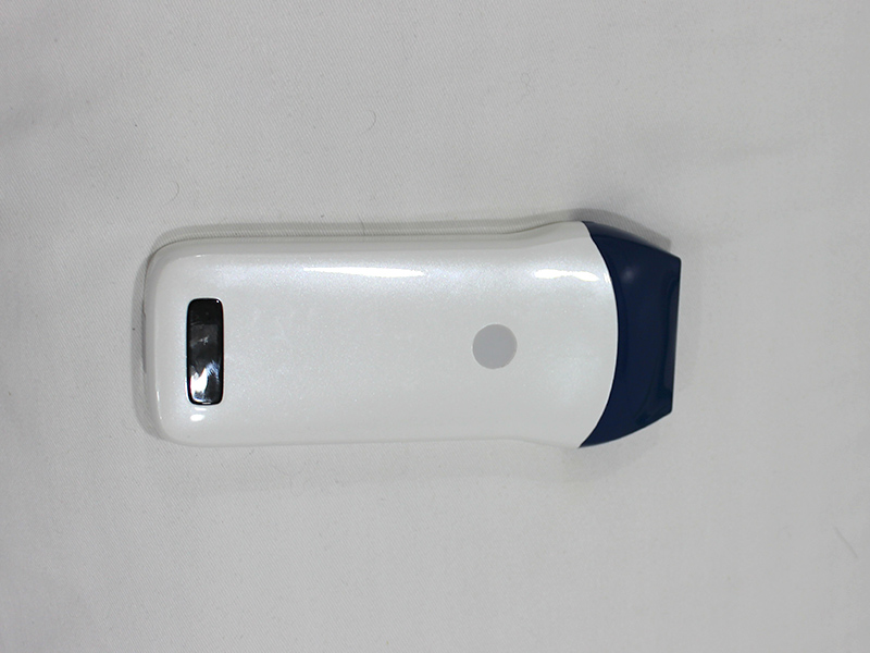
Probe Physical Specs: The linear probe adopts an ergonomic flat-face design, ensuring good skin contact even in awkward anatomical areas. It matches the scanner’s 250g weight distribution, enabling comfortable handheld operation during prolonged clinical examinations.
2. Imaging Quality & Optimization Functions
Comprehensive imaging modes and adjustment tools to enhance superficial imaging clarity and diagnostic precision:
-
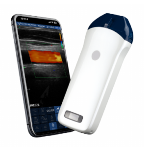
Imaging Modes: Supports B, B/M, Color, PDI (Power Doppler Imaging), and PW (Pulsed Wave Doppler) modes. It covers static tissue morphology observation, dynamic process monitoring (e.g., vascular pulsation, muscle contraction), and blood flow detection, providing multi-dimensional diagnostic information for superficial tissues.
-
Imaging Foundation: 256-level image gray scale accurately reproduces subtle density differences between normal tissues and lesions, laying a solid foundation for precise identification of tiny superficial abnormalities.
-
Image Adjustment: Equipped with comprehensive optimization tools, including Gain (30dB-105dB), 8-level Dynamic Range (40-110), and 5-level Noise Reduction (0-4). These tools effectively reduce image noise, balance tissue brightness across different depths, and enhance image layering, further improving the clarity of superficial tissue imaging.
3. Clinical Functions & Data Management
Practical clinical tools and flexible data handling to improve superficial tissue examination efficiency:
-
Measurement Functions: Supports distance, area, obstetrics, and other clinical measurements, providing accurate quantitative data. It assists in evaluating lesion size, tissue thickness, and obstetric indicators, laying a reliable foundation for clinical decision-making and follow-up assessments.
-
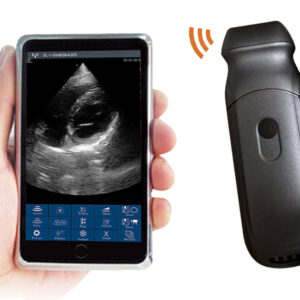
Data Storage: Images and videos are directly stored on mobile phones or tablets, eliminating the need for additional storage devices. This design facilitates on-site review of high-resolution images, quick case sharing with colleagues, and easy data archiving for follow-up comparison and medical records.
4. System Compatibility & Physical Specifications
Cross-system compatibility and portable design optimized for mobile clinical scenarios:
-
Display & Compatibility: Adapts to smartphone or tablet screens; compatible with iOS, Android, and Windows operating systems, enabling flexible connection to various terminal devices. This cross-system design reduces terminal replacement costs and adapts to different clinical usage habits of medical professionals.
-
Power Supply: Equipped with a built-in 4200mAh lithium battery, supporting 3 hours of continuous operation, fully covering daily clinical examination needs without frequent charging, ideal for mobile or bedside examinations.
-
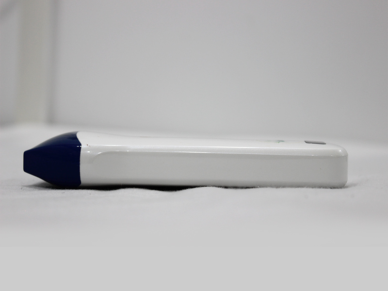
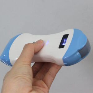
Physical Dimensions: 156mm×60mm×20mm compact size, 250g net weight, with an ergonomic handheld design for comfortable one-hand operation. It can be easily carried in a medical bag, suitable for bedside examinations, home visits, community healthcare, and field medical services.
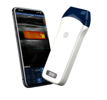
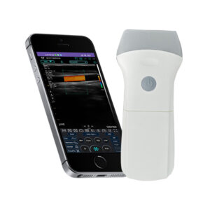
The VISION5 L linear palm Doppler ultrasound scanner integrates 7.5/10MHz dual-frequency imaging, 128-element signal processing, and an expanded L40 field of view. It provides stable and precise superficial tissue imaging solutions for diverse clinical scenarios, balancing diagnostic versatility and operational flexibility. As a reliable tool for medical professionals, it meets the demands of routine examinations and specialized assessments in primary healthcare and clinical settings.
Your message has been sent

Reviews
There are no reviews yet.