
Description
1. Core Scanning & High-Frequency Probe Parameters
Foundational hardware that defines ultra-precise superficial imaging:
- Scanning & Probe Design: Electronic Array Linear scanning, equipped with a high-frequency dual-band probe (18/22MHz) optimized for ultra-superficial tissues. The frequency configuration is tailored for specialized needs: 18MHz balances resolution and penetration for small organs (e.g., thyroid nodules, parotid glands) and micro-vascular networks, while 22MHz delivers near-microscopic clarity for delicate structures (e.g., peripheral nerve bundles, skin layers, pediatric superficial tissues).
- Signal Processing: 192 high-precision elements and 64 dedicated channels ensure exceptional signal-to-noise ratio, minimizing image distortion and enhancing the definition of tissue boundaries. This advanced signal processing is critical for detecting sub-millimeter lesions, subtle anatomical variations, and micro-vascular abnormalities that may be invisible to lower-frequency scanners.
- Imaging Range: Adjustable scanning depth (10/15/20/25mm) tailored to ultra-superficial tissue layers, covering the full spectrum of superficial anatomical targets. An 18mm field of view provides focused, high-density coverage, concentrating on target areas to eliminate background clutter and maximize detail extraction.
2. Imaging Quality & Optimization Functions
Comprehensive tools to maximize clarity for high-frequency imaging:
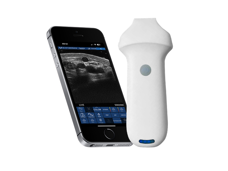
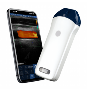
- Imaging Modes & Foundation: Supports B, B/M, Color, PDI (Power Doppler Imaging), and PW (Pulsed Wave Doppler) modes. 256-level image gray scale accurately reproduces subtle tissue density differences, enabling precise differentiation between healthy and abnormal tissues—essential for diagnosis in specialized fields like dermatology, neuromuscular medicine, and pediatric radiology.
- Image Adjustment Suite: Equipped with multi-dimensional optimization features, including Gain (30dB-105dB), Focus, Reverse Pulse Harmonics, and 5-level Noise Reduction (0-4). These tools allow operators to fine-tune image brightness, suppress background interference, and enhance edge definition, adapting to the unique properties of ultra-superficial tissues (e.g., thin skin layers, fragile nerve fibers, tiny blood vessels) for optimal visualization.
- Dynamic Range: 8-level adjustable dynamic range (40-110) enables customization of image contrast, ensuring clear differentiation of tissue components even in dense or complex superficial structures.
3. Clinical Functions & Data Management
Specialized features tailored to interventional and diagnostic workflows:
- Puncture Assistance: In-Plane/Out-Plane puncture guide lines and automatic vascular measurement function support precise interventional procedures (e.g., micro-vascular cannulation, nerve block guidance, small lesion biopsy). These features reduce procedural time, minimize patient discomfort, and improve targeting accuracy for minimally invasive interventions.
- Measurement & Cineloop: Supports precise quantitative measurements (distance, area, obstetrics) for evaluating lesion size, tissue thickness, and vascular diameter—vital for diagnosis, treatment planning, and follow-up. Manual/automatic Cineloop with adjustable playback frames (100/200/500/1000) enables frame-by-frame review of dynamic processes (e.g., micro-vascular blood flow, nerve movement).
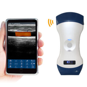
- Data Storage & Connectivity: Images/videos are stored in JPG, PNG, MP4, and DCM formats directly on mobile devices or tablets, ensuring compatibility with electronic medical records (EMRs) and hospital PACS systems. WiFi 802.11n/2.4G/5G dual-band (450Mbps) enables fast wireless data transfer and remote consultation with colleagues.
4. Power & Physical Specifications
Portability and endurance optimized for specialized use:
- Power Supply: Built-in 2200mAh lithium battery with two versions—regular (1.5 hours of continuous use) for short shifts or quick examinations, and larger battery version (3 hours) for extended clinical work or research sessions. Wireless charging support extends operation indefinitely, ideal for prolonged procedures or field medical services without access to power outlets.
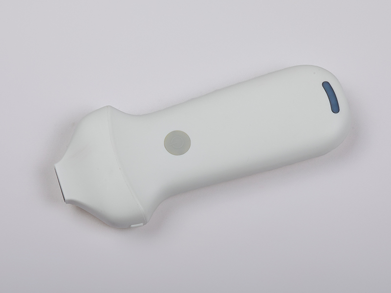
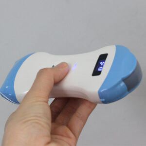
- Ultra-Compact Design: Dimensions of 156mm×60mm×20mm and 180g net weight make it highly portable, fitting easily in a medical bag or pocket. Its ergonomic handheld design reduces operator fatigue during prolonged use, while durable construction withstands the demands of daily clinical and research environments.
- System Compatibility: Seamlessly works with iOS, Android, and Windows operating systems, supporting flexible connection to smartphones, tablets, and computers. This cross-platform adaptability ensures compatibility with existing clinical setups and user preferences, enhancing its practicality across diverse healthcare settings.
The VISION8 S Linear Palm Doppler Ultrasound Scanner sets a new standard for high-frequency superficial imaging, combining microscopic resolution, specialized clinical functionality, and user-centric portability. It is an indispensable tool for dermatologists, neuromuscular specialists, pediatricians, interventional radiologists, and researchers seeking precise, efficient imaging of ultra-superficial structures. Whether in a specialized clinic, research lab, or remote field setting, it delivers consistent, high-quality results to support timely and accurate clinical decisions, redefining the possibilities of palm-sized ultrasound in specialized medicine.

Reviews
There are no reviews yet.